ARTICLES

Hyperfine splitting and ferromagnetism in CdS : Mn nanoparticles for optoelectronic device applications

Madhavi Sharad Darekar and Praveen Beekanahalli Mokshanatha

+ Author Affiliations

 Corresponding author: Madhavi Sharad Darekar, madhavi_darekar28@rediff.com

PDF

Turn off MathJax

Abstract: Manganese (Mn) doped cadmium sulphide (CdS) nanoparticles were synthesized using a chemical method. It was possible to decrease CdS : Mn particle size by increasing Mn concentration. Investigation techniques such as ultraviolet−visible (UV−Vis) absorption spectroscopy and photoluminescence (PL) spectroscopy were used to determine optical properties of CdS : Mn nanoparticles. Size quantization effect was observed in UV−Vis absorption spectra. Quantum efficiency for luminescence or the internal magnetic field strength was increased by doping CdS nanoparticles with Mn element. Orange emission was observed at wavelength ~630 nm due to 4T16A1 transition. Isolated Mn2+ ions arranged in tetrahedral coordination are mainly responsible for luminescence. Luminescence quenching and the effect of Mn doping on hyperfine interactions in the case of CdS nanoparticles were also discussed. The corresponding weight percentage of Mn element actually incorporated in doping process was determined by atomic absorption spectroscopy (AAS). Crystallinity was checked and the average size of nanoparticles was estimated using the X-ray diffraction (XRD) technique. CdS : Mn nanoparticles show ferromagnetism at room temperature. Transmission electron microscopy (TEM) images show spherical clusters of various sizes and selected area electron diffraction (SAED) patterns show the polycrystalline nature of the clusters. The electronic states of diluted magnetic semiconductors (DMS) of Ⅱ−Ⅵ group CdS nanoparticles give them great potential for applications due to quantum confinement. In this study, experimental results and discussions on these aspects have been given.

Key words: synthesischaracterizationundoped CdS nanoparticlesMn-doped CdS nanoparticlesnon-aqueous chemical method


[1]
Gadalla A, Almokhtar M, Abouelkhir A N. Effect of Mn doping on structural, optical and magnetic properties of CdS diluted magnetic semiconductor nanoparticles. Chalcogenide Letters, 2018, 15, 207
[2]
Chaure S. Investigation of the effect of manganese doping in CdS nanocrystalline thin films. Mater Res Express, 2018, 6, 025912 doi: 10.1088/2053-1591/aad4e1
[3]
Venkatesan D, Deepan D, Ramkumar J, et al. Synthesis and characterization of sodium bis (2-ethylhexyl) sulfonsuccinate (AOT) capped pure and Mn-doped CdS nanoparticles. J Nanomater, 2012, 2012, 1 doi: 10.1155/2012/492573
[4]
Maity P, Kumar R, Jha S N, et al. Investigation of the Physical Properties of Mn-doped CdS Diluted Magnetic Semiconductor Quantum Dots: Non-linear Band Gap variation with Downwards Bowing. Available at SSRN 4066164
[5]
Levy L, Ingert D, Feltin N, et al. Cd1− y Mn y S nanoparticles: Absorption and photoluminescence properties. J Cryst Growth, 1998, 184/185, 377 doi: 10.1016/S0022-0248(97)00780-X
[6]
Hofmann D M, Hofstaetter A, Leib U, et al. EPR and ENDOR investigations on CdS : Mn nanocrystals. J Cryst Growth, 1998, 184/185, 383 doi: 10.1016/S0022-0248(98)80081-X
[7]
Counio G, Esnouf S, Gacoin T, et al. CdS : Mn nanocrystals in transparent xerogel matrices: synthesis and luminescence properties. J Phys Chem, 1996, 100, 20021 doi: 10.1021/jp961937i
[8]
Bhargava R N, Gallagher D, Hong X, et al. Optical properties of manganese-doped nanocrystals of ZnS. Phys Rev Lett, 1994, 72, 416 doi: 10.1103/PhysRevLett.72.416
[9]
Khosravi A A, Kundu M, Jatwa L, et al. Green luminescence from copper doped zinc sulphide quantum particles. Appl Phys Lett, 1995, 67, 2702 doi: 10.1063/1.114298
[10]
Khosravi A A, Kundu M, Kuruvilla B A, et al. Manganese doped zinc sulphide nanoparticles by aqueous method. Appl Phys Lett, 1995, 67, 2506 doi: 10.1063/1.114440
[11]
Bhargava R N, Gallagher D, Welker T, et al. Doped nanocrystals of semiconductors - a new class of luminescent materials. J Lumin, 1994, 60/61, 275 doi: 10.1016/0022-2313(94)90146-5
[12]
Bhargava R N. Nanoparticles and their use for multifunctional bioimaging. J Lumin, 1996, 70, 85 doi: 10.1016/0022-2313(96)00046-4
[13]
Norris D J, Yao N, Charnock F T, et al. High-quality manganese-doped ZnSe nanocrystals. Nano Lett, 2001, 1, 3 doi: 10.1021/nl005503h
[14]
Ochsenbein S T, Gamelin D R. Quantum oscillations in magnetically doped colloidal nanocrystals. Nat Nanotechnol, 2011, 6, 112 doi: 10.1038/nnano.2010.252
[15]
Beaulac R, Ochsenbein S, Gamelin D, et al. Colloidal transition-metal-doped quantum dots. In nanocrystal quantum dots. CRC Press, 2010, 397
[16]
Yu J H, Liu X Y, Kweon K E, et al. Giant Zeeman splitting in nucleation-controlled doped CdSe : Mn2+ quantum nanoribbons. Nat Mater, 2010, 9, 47 doi: 10.1038/nmat2572
[17]
Wood V, Halpert J E, Panzer M J, et al. Alternating Current driven electroluminescence from ZnSe/ZnS : Mn/ZnS nanocrystals. Nano Lett, 2009, 9, 2367 doi: 10.1021/nl900898t
[18]
Levy L, Feltin N, Ingert D, et al. Three dimensionally diluted magnetic semiconductor clusters Cd1- y Mn y S with a range of sizes and compositions: dependence of spectroscopic properties on the synthesis mode. J Phys Chem B, 1997, 101, 9153 doi: 10.1021/jp970978r
[19]
Liu S M, Liu F Q, Guo H Q, et al. Surface states induced photoluminescence from Mn2+ doped CdS nanoparticles. Solid State Commun, 2000, 115, 615 doi: 10.1016/S0038-1098(00)00254-4
[20]
McClure D S. Electronic spectra of molecules and ions in crystals part II. Solid State Physics, 1959, 9, 399 doi: 10.1016/S0081-1947(08)60569-X
[21]
Tanabe Y, Sugano S. On the absorption spectra of complex ions I, II. J Phys Soc Jpn, 1954, 9, 753 doi: 10.1143/JPSJ.9.753
[22]
Levy L, Hochepied J F, Pileni M P. Control of the size and composition of three dimensionally diluted magnetic semiconductor clusters. J Phys Chem, 1996, 100, 18322 doi: 10.1021/jp960824w
[23]
Romčević N, Kostic R, Romčević M, et al. Raman spectroscopy of Cd1− x Mn x S quantum dots. J Phys D: Appl Phys, 2005, 38, 4321 doi: 10.1088/0022-3727/38/24/009
[24]
Patle U S, Ahirwar R K, Bhatt A, et al. Enhanced photoluminescence properties of Mn doped CdS nanocrystals. AIP Conf Proc, 2019, 2100, 020165-1 doi: 10.1063/1.5098719
[25]
Babi Stoji B, Milivojevi D, Comor M, et al. Optical and electron paramagnetic resonance spectroscopy of Cd1- x Mn x S quantum dots. J Phys: Condens Matter, 2004, 16, 4625 doi: 10.1088/0953-8984/16/25/020
[26]
Patel N H, Deshpande M P, Bhatt S V, et al. Structural and magnetic properties of undoped and Mn doped CdS nanoparticles prepared by chemical co-precipitation method. Adv Mater Lett, 2014, 5, 671 doi: 10.5185/amlett.2014.1574
[27]
Chauhan R, Kumar A, Chaudhary R P. Synthesis, structural and photocatalytic studies of Mn-doped CdS nanoparticles. Res Chem Intermed, 2013, 39, 645 doi: 10.1007/s11164-012-0586-x
[28]
Gadalla A A, Aboelkhir A N, Mahesha M G, et al. RETRACTED ARTICLE : Synthesis and characterization of Mn-doped CdS-diluted magnetic semiconductor nanoparticles. J Mater Sci: Mater Electron, 2020, 31, 10941 doi: 10.1007/s10854-020-03240-x
[29]
Venkatesu P. Doping effect of Mn on structural optical magnetic and electrical properties of CdS nanoparticles. International Conference on Advanced Nanomaterials & Emerging Engineering Technologies, 2013, 260 doi: 10.1109/ICANMEET.2013.6609255
[30]
Malik M A, O' Brien P, Revaprasadu N. Synthesis of TOPO-capped Mn-doped ZnS and CdS quantum dots. J Mater Chem, 2001, 11, 2382 doi: 10.1039/b102709n
[31]
Keerthana S, Yuvakkumar R, Ravi G, et al. PVP influence on Mn-CdS for efficient photocatalytic activity. Chemosphere, 2021, 277, 130346 doi: 10.1016/j.chemosphere.2021.130346
[32]
Patle U S. Synthesis and characterization of Mn doped CdS nanoparticles prepared by chemical bath deposition method. International Journal of Science and Research (IJSR), 2015, 4, 1945
[33]
Darekar M S, Praveen B M. Synthesis and characterization of nanoparticles of semiconducting metal suplhide and their application. Phys Scr, 2022, 97, 065805 doi: 10.1088/1402-4896/ac698f
[34]
Darekar M S, Praveen B M. High photosensitivity nanocrystalline p-Cu2S/n-FTO heterojunction photodetectors prepared by dip coating method. J Mod Nanotechnol, 2023, 3 doi: 10.53964/jmn.2023001
[35]
Dareka M S, Praveen B M. Effects of heat treatment in air atmosphere on dip coating deposited CdS thin films for photo sensor applications. J Mod Nanotechnol, 2023, 3 doi: 10.53964/jmn.2023002
[36]
Ikeda M, Itoh K, Sato H. Electrical and optical properties of CdS-MnS single crystals. J Phys Soc Jpn, 1968, 25, 455 doi: 10.1143/JPSJ.25.455
[37]
Tsai C T, Chen S H, Chuu D S, et al. Fabrication and physical properties of radio frequency sputtered Cd1– x Mn x S thin films. Phys Rev B, 1996, 54, 11555 doi: 10.1103/PhysRevB.54.11555
[38]
Costa V C, Shen Y R, Bray K L. Luminescence properties of nanocrystalline CdS and CdS : Mn2+ doped silica-type glasses. J Non Cryst Solids, 2002, 304, 217 doi: 10.1016/S0022-3093(02)01026-8
[39]
Hoffman D M, Meyer B K, Ekimov A I, et al. Giant internal magnetic fields in Mn doped nanocrystal quantum dots. Solid State Commun, 2000, 114, 547 doi: 10.1016/S0038-1098(00)00089-2
[40]
Zhang Y N, Raman N, Bailey J K, et al. A new sol-gel route for the preparation of nanometer-scale semiconductor particles that exhibit quantum optical behavior. J Phys Chem, 1992, 96, 9098 doi: 10.1021/j100202a004
[41]
Oka Y, Yanata K. Excitonic properties of nanostructure semimagnetic semiconductors. J Lumin, 1996, 70, 35 doi: 10.1016/0022-2313(96)82859-6
[42]
de Mello Donegá C, Bol A A, Meijerink A. Time-resolved luminescence of ZnS : Mn2+ nanocrystals. J Lumin, 2002, 96, 87 doi: 10.1016/S0022-2313(01)00418-5
[43]
Ethiraj A S, Hebalkar N, Kulkarni S K, et al. Enhancement of photoluminescence in manganese-doped ZnS nanoparticles due to a silica shell. J Chem Phys, 2003, 118, 8945 doi: 10.1063/1.1566932
[44]
Samarth N, Furdyna J K. Electron paramagnetic resonance in Cd1- x Mn x S, Cd1- x Mn x Se, and Cd1- x Mn x Te. Phys Rev B Condens Matter, 1988, 37, 9227 doi: 10.1103/PhysRevB.37.9227
[45]
Venkatesu P, Ravichandran K. Manganese doped cadmium sulphide (CdS : Mn) quantum particles: Topological, photoluminescence and magnetic studies. Adv Mater Lett, 2013, 4, 202 doi: 10.5185/amlett.2012.7379
[46]
Maaz K, Karim S, Lee K J, et al. Effect of temperature on the magnetic characteristics of Ni0.5Co0.5Fe2O4 nanoparticles. Mater Chem Phys, 2012, 133, 1006 doi: 10.1016/j.matchemphys.2012.02.007
[47]
Feltin N, Levy L, Ingert D, et al. Magnetic properties of 4-nm Cd1- y Mn y S nanoparticles differing by their compositions, y. J Phys Chem B, 1999, 103, 4 doi: 10.1021/jp981241k
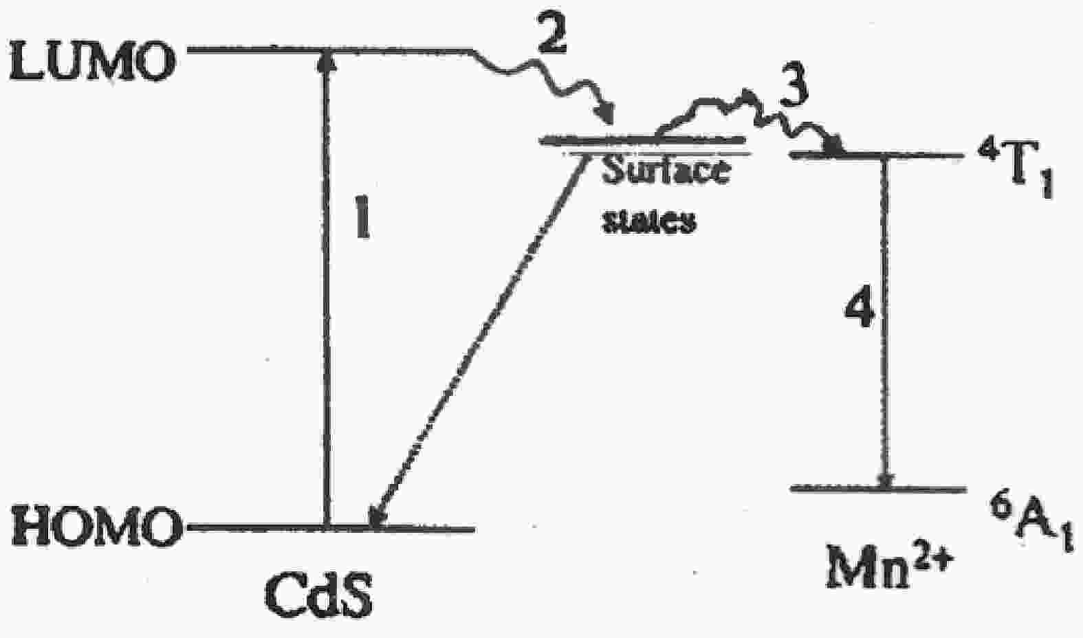
Fig. 1.  Luminescence mechanism scheme of Mn-doped CdS nanoparticles[19].

Fig. 2.  UV−Vis absorption spectra of Mn-doped CdS nanoparticles (samples A to J).

Fig. 3.  Variation of energy gap with atomic weight percentage of Mn in Mn-doped CdS nanoparticles (samples A to J).

Fig. 4.  UV−Vis absorption spectrum of Mn-doped CdS nanoparticles (sample K).

Fig. 5.  UV−Vis absorption spectra of Mn-doped CdS nanoparticles (samples R to Z).

Fig. 6.  Variation of energy gap with particle size in Mn-doped CdS nanoparticles (samples R to Z).

Fig. 7.  UV−Vis absorption spectra of Mn-doped CdS nanoparticles (samples A’, B’, C’ and D’).

Fig. 8.  XRD spectra of Mn-doped CdS nanoparticles (samples A to J).

Fig. 9.  Excitation and emission spectra of Mn-doped CdS nanoparticles (samples A to J).

Fig. 10.  Variation of PL intensity with atomic weight percentage of Mn in samples A to J.

Fig. 11.  ESR spectra of Mn-doped CdS nanoparticles (samples A to J).

Fig. 12.  Variation of line width with atomic weight percentage of Mn (samples A to J).

Fig. 13.  ESR spectra of manganese sulphide nanoparticles.

Fig. 14.  ESR spectra of Mn-doped CdS nanoparticles (samples R to Z).

Fig. 15.  Variation of line width with weight percentage of Mn in samples R to Z.

Fig. 16.  ESR spectra of Mn-doped CdS nanoparticles (samples A’, B’, C’ and D’).

Fig. 17.  Variation of line width with atomic weight percentage of Mn in samples A’, B’, C’, and D’.

Fig. 18.  M(H) curves of two Cd1 – xMnxS samples (x = 0.04, 0.05) at 300 K.

Fig. 19.  Temperature dependence of magnetization for Cd0.96Mn0.04S at 1 KOe. The inset shows the best fit to the Bloch law M(T) = M(0) × (1 − AT3/2) (shown by the marked line).

Fig. 20.  Total energy difference ∆EFM-AFM between ferromagnetic and anti-ferromagnetic configurations for Mn-doped CdS nanoparticles as a function of Mn concentration in the “short” (squares) and “far” (circles) configurations.

Fig. 21.  (a) TEM image of Mn-doped CdS nanoparticles (sample A) with magnification of 200 nm and (b) SAED pattern of Mn-doped CdS nanoparticles (sample A).

Fig. 22.  (a) TEM image of Mn-doped CdS nanoparticles (sample J) with magnification of 200 nm and (b) SAED pattern of Mn-doped CdS nanoparticles (sample J).

Table 1.   Properties of thioglycerol.

ParameterSHCH2CH(OH)CH2OH
Density1.295 gm/mL
Molecular weight108.16 gm/mole
Purity99%
Molarity11.9 M
Boiling point118 °C
Freezing point110 °C
DownLoad: CSV

Table 2.   UV−Vis absorption values, band gap values and particle sizes of Mn-doped CdS nanoparticles (samples A to J).

Sample Wt.% of Mn used for chemical reaction Wt.% of Mn found by AAS At. Wt.% of Mn found by AAS UV−Vis absorp-
tion (nm)
Band gap
Eg (eV)
Particle size (nm)
A 15 3.72 0.068 358 3.46 5.28
B 25 4.56 0.083 365 3.40 5.36
C 35 5.66 0.103 350 3.54 5.18
D 45 6.48 0.118 340 3.65 5.06
E 50 6.87 0.125 336 3.69 5.02
F 55 7.20 0.131 330 3.76 4.95
G 60 7.53 0.137 318 3.90 4.81
H 65 7.80 0.142 315 3.94 4.78
I 70 8.02 0.146 310 4.00 4.73
J 80 8.46 0.154 300 4.14 4.61
DownLoad: CSV

Table 3.   UV−Vis absorption values, band gap values and particle sizes of Mn-doped CdS nanoparticles (samples R to Z).

SampleWt.% of Mn used for
chemical reaction
Wt.% of Mn found by
AAS
At. Wt.% of Mn found by AASUV−Vis absorption (nm)Band gap Eg (eV)Particle size (nm)
R6.4 × 10−50.026 ± 0.0020.00053463.595.13
S8 × 10−50.0780.00143503.545.18
T2 × 10−40.1320.00243503.545.18
U3 × 10−40.1520.00283523.525.21
V4 × 10−40.240.00443553.495.24
W5 × 10−40.30.00553573.475.27
X7 × 10−40.620.01133603.455.29
Y1 × 10−31.240.02263663.395.37
Z5 × 10−33.720.06713703.355.42
DownLoad: CSV

Table 4.   UV−Vis absorption values, band gap values and particle sizes of Mn-doped CdS nanoparticles (samples A’, B’, C’, and D’).

Sample At. Wt.% of Mn found by AAS UV−Vis absorption (nm) Band gap Eg (eV) Particle size (nm)
A’ 0.0080 370 3.35 5.42
B’ 0.0085 350 3.54 5.18
C’ 0.0176 336 3.69 5.02
D’ 0.0602 320 3.88 4.83
DownLoad: CSV

Table 5.   XRD parameters of Mn-doped CdS nanoparticles (samples A to J).

Sample Wt.% of Mn used for chemical reaction Wt.% of Mn found
by AAS
At. Wt.% of Mn found by AAS XRD average particle size, d’ (nm) and inter planar distance, d (Å)
A 15 3.72 0.068 2$ \theta $ = 29°
d’ = 5.27 nm
d = 3.08 Å
2$ \theta $ = 47.8°
d’ = 3.72 nm
d = 1.90 Å
B 25 4.56 0.083 2$ \theta $ = 28.6°
d’ = 5.70 nm
d = 3.12 Å
2$ \theta $ = 47.7°
d’ = 3.81 nm
d = 1.90 Å
C 35 5.66 0.103 2$ \theta $ = 28.7°
d’ = 5.33 nm
d = 3.11 Å
2$ \theta $ = 47.5°
d’ = 3.62 nm
d = 1.91 Å
D 45 6.48 0.118 2$ \theta $ = 29.2°
d’ = 4.89nm
d = 3.05 Å
2$ \theta $ = 47.6°
d’ = 4.58 nm
d = 1.91 Å
E 50 6.87 0.125 2$ \theta $ = 28.6°
d’ = 5.19 nm
d = 3.12 Å
2$ \theta $ = 47.4°
d’ = 4.28 nm
d = 1.92 Å
F 55 7.20 0.131 2$ \theta $ = 28.8°
d’ = 4.89 nm
d = 3.1 Å
2$ \theta $ = 47.3°
d’ = 3.56 nm
d = 1.92 Å
G 60 7.53 0.137 2$ \theta $ = 28.5°
d’ = 5.4 nm
d = 3.13 Å
2$ \theta $ = 47.5°
d’ = 3.81 nm
d = 1.91 Å
H 65 7.80 0.142 2$ \theta $ = 28.6°
d’ = 4.94 nm
d = 3.12 Å
2$ \theta $ = 47.2°
d’ = 3.84 nm
d = 1.92 Å
I 70 8.02 0.146 2$ \theta $ = 28.8°
d’ = 3.73 nm
d = 3.1 Å
2$ \theta $ = 47.1°
d’ = 2.83 nm
d = 1.93 Å
J 80 8.46 0.154 2$ \theta $ = 28.7°
d’ = 4.77 nm
d = 3.11 Å
2$ \theta $ = 47.7°
d’ = 3.53 nm
d = 1.90 Å
DownLoad: CSV

Table 6.   PL parameters of Mn-doped CdS nanoparticles (samples A to J).

Sample Wt.% of Mn used for chemical reaction Wt.% of Mn found by AAS At. Wt.% of Mn found by AAS
PL intensity
Excitation
λex (nm)
Emission λem (nm)
A 15 3.72 0.068 391.3 625
B 25 4.56 0.083 389.2 626
C 35 5.66 0.103 393.1 628
D 45 6.48 0.118 392.9 631
E 50 6.87 0.125 391.9 634
F 55 7.20 0.131 391.3 638
G 60 7.53 0.137 389 634
H 65 7.80 0.142 387.9 633
I 70 8.02 0.146 389 642
J 80 8.46 0.154 386.1 631
DownLoad: CSV

Table 7.   ESR parameters of Mn-doped CdS nanoparticles (samples A to J).

Sample At. Wt.% of Mn Line width
(Gauss)
Spectroscopic splitting factor (g) Bra
(Gauss)
ESR intensity (× 105) ESR intensity/gain No. of CdS molecules No. of Mn atoms
A 0.068 225.69 2.0079 3465.8418 1.9101743 1.9101743 6909 1008
B 0.083 243.05 2.0079 3462.2744 4.7703872 1.1925968 4078 628
C 0.103 294.11 2.0128 3457.3715 4.4982699 4.4982699 8038 1053
D 0.118 302.76 2.0077 3466.022 2.7958911 2.7958911 4754 561
E 0.125 302.76 2.0077 3466.022 3.7125768 3.7125768 1998 250
F 0.131 302.76 2.0178 3445.1535 8.6626791 5.4141 6582 934
G 0.137 328.71 2.0178 3448.7209 5.6189461 3.5118413 6625 908
H 0.142 397.92 2.0178 3448.7209 5.0669891 5.0669891 3897 401
I 0.146 501.73 2.0028 3474.6725 2.4355177 19.4841 5231 434
J 0.154 527.68 1.9854 3504.9493 2.561721 16.010756 5384 307
a: Br is the magnetic field at the center of the two resonance lines.
DownLoad: CSV

Table 8.   ESR parameters of Mn-doped CdS nanoparticles (samples R to Z).

Sample At. Wt.% of Mn found by AAS Spectroscopic splitting factor (g) Line width (Gauss)
R 0.0005 1.9904 10
S 0.0014 1.9904 19.16
T 0.0024 1.9910 54.17
U 0.0028 1.9902 55
V 0.0044 1.9902 55.87
W 0.0055 1.9902 56.7
X 0.0113 1.9910 59.2
Y 0.0226 1.9909 60
Z 0.0671 1.9904 80.8
DownLoad: CSV

Table 9.   ESR parameters of Mn-doped CdS nanoparticles (samples A’, B’, C’, and D’).

Sample Wt.% of Mn found by AAS At. Wt.% of Mn found by AAS Line width (Gauss) Br (Gauss) ESR intensity
(×105)
Spectroscopic splitting factor (g)
A’ 0.439 0.0080 97.7 3370.5 0.1055 2.068
B’ 0.469 0.0085 101.7 2535.7 2.5 2.050
C’ 0.969 0.0176 144.7 3374.4 0.955 2.066
D’ 3.309 0.0602 164.2 3384.2 1.087 2.060
DownLoad: CSV

Table 10.   VSM parameters of two Cd1 – xMnxS powder samples (x = 0.04, 0.05).

SampleMs at 300 K
(emu/g)
Ms at 5 K
(emu/g)
Hc at 300 K
(Oe)
Hc at 5 K
(Oe)
Cd0.95Mn0.05S~1.61 × 10⁻³~1.59 × 10⁻²~105~250
Cd0.96Mn0.04S~3.07 × 10⁻³~3.84 × 10⁻²~100~98
DownLoad: CSV
[1]
Gadalla A, Almokhtar M, Abouelkhir A N. Effect of Mn doping on structural, optical and magnetic properties of CdS diluted magnetic semiconductor nanoparticles. Chalcogenide Letters, 2018, 15, 207
[2]
Chaure S. Investigation of the effect of manganese doping in CdS nanocrystalline thin films. Mater Res Express, 2018, 6, 025912 doi: 10.1088/2053-1591/aad4e1
[3]
Venkatesan D, Deepan D, Ramkumar J, et al. Synthesis and characterization of sodium bis (2-ethylhexyl) sulfonsuccinate (AOT) capped pure and Mn-doped CdS nanoparticles. J Nanomater, 2012, 2012, 1 doi: 10.1155/2012/492573
[4]
Maity P, Kumar R, Jha S N, et al. Investigation of the Physical Properties of Mn-doped CdS Diluted Magnetic Semiconductor Quantum Dots: Non-linear Band Gap variation with Downwards Bowing. Available at SSRN 4066164
[5]
Levy L, Ingert D, Feltin N, et al. Cd1− y Mn y S nanoparticles: Absorption and photoluminescence properties. J Cryst Growth, 1998, 184/185, 377 doi: 10.1016/S0022-0248(97)00780-X
[6]
Hofmann D M, Hofstaetter A, Leib U, et al. EPR and ENDOR investigations on CdS : Mn nanocrystals. J Cryst Growth, 1998, 184/185, 383 doi: 10.1016/S0022-0248(98)80081-X
[7]
Counio G, Esnouf S, Gacoin T, et al. CdS : Mn nanocrystals in transparent xerogel matrices: synthesis and luminescence properties. J Phys Chem, 1996, 100, 20021 doi: 10.1021/jp961937i
[8]
Bhargava R N, Gallagher D, Hong X, et al. Optical properties of manganese-doped nanocrystals of ZnS. Phys Rev Lett, 1994, 72, 416 doi: 10.1103/PhysRevLett.72.416
[9]
Khosravi A A, Kundu M, Jatwa L, et al. Green luminescence from copper doped zinc sulphide quantum particles. Appl Phys Lett, 1995, 67, 2702 doi: 10.1063/1.114298
[10]
Khosravi A A, Kundu M, Kuruvilla B A, et al. Manganese doped zinc sulphide nanoparticles by aqueous method. Appl Phys Lett, 1995, 67, 2506 doi: 10.1063/1.114440
[11]
Bhargava R N, Gallagher D, Welker T, et al. Doped nanocrystals of semiconductors - a new class of luminescent materials. J Lumin, 1994, 60/61, 275 doi: 10.1016/0022-2313(94)90146-5
[12]
Bhargava R N. Nanoparticles and their use for multifunctional bioimaging. J Lumin, 1996, 70, 85 doi: 10.1016/0022-2313(96)00046-4
[13]
Norris D J, Yao N, Charnock F T, et al. High-quality manganese-doped ZnSe nanocrystals. Nano Lett, 2001, 1, 3 doi: 10.1021/nl005503h
[14]
Ochsenbein S T, Gamelin D R. Quantum oscillations in magnetically doped colloidal nanocrystals. Nat Nanotechnol, 2011, 6, 112 doi: 10.1038/nnano.2010.252
[15]
Beaulac R, Ochsenbein S, Gamelin D, et al. Colloidal transition-metal-doped quantum dots. In nanocrystal quantum dots. CRC Press, 2010, 397
[16]
Yu J H, Liu X Y, Kweon K E, et al. Giant Zeeman splitting in nucleation-controlled doped CdSe : Mn2+ quantum nanoribbons. Nat Mater, 2010, 9, 47 doi: 10.1038/nmat2572
[17]
Wood V, Halpert J E, Panzer M J, et al. Alternating Current driven electroluminescence from ZnSe/ZnS : Mn/ZnS nanocrystals. Nano Lett, 2009, 9, 2367 doi: 10.1021/nl900898t
[18]
Levy L, Feltin N, Ingert D, et al. Three dimensionally diluted magnetic semiconductor clusters Cd1- y Mn y S with a range of sizes and compositions: dependence of spectroscopic properties on the synthesis mode. J Phys Chem B, 1997, 101, 9153 doi: 10.1021/jp970978r
[19]
Liu S M, Liu F Q, Guo H Q, et al. Surface states induced photoluminescence from Mn2+ doped CdS nanoparticles. Solid State Commun, 2000, 115, 615 doi: 10.1016/S0038-1098(00)00254-4
[20]
McClure D S. Electronic spectra of molecules and ions in crystals part II. Solid State Physics, 1959, 9, 399 doi: 10.1016/S0081-1947(08)60569-X
[21]
Tanabe Y, Sugano S. On the absorption spectra of complex ions I, II. J Phys Soc Jpn, 1954, 9, 753 doi: 10.1143/JPSJ.9.753
[22]
Levy L, Hochepied J F, Pileni M P. Control of the size and composition of three dimensionally diluted magnetic semiconductor clusters. J Phys Chem, 1996, 100, 18322 doi: 10.1021/jp960824w
[23]
Romčević N, Kostic R, Romčević M, et al. Raman spectroscopy of Cd1− x Mn x S quantum dots. J Phys D: Appl Phys, 2005, 38, 4321 doi: 10.1088/0022-3727/38/24/009
[24]
Patle U S, Ahirwar R K, Bhatt A, et al. Enhanced photoluminescence properties of Mn doped CdS nanocrystals. AIP Conf Proc, 2019, 2100, 020165-1 doi: 10.1063/1.5098719
[25]
Babi Stoji B, Milivojevi D, Comor M, et al. Optical and electron paramagnetic resonance spectroscopy of Cd1- x Mn x S quantum dots. J Phys: Condens Matter, 2004, 16, 4625 doi: 10.1088/0953-8984/16/25/020
[26]
Patel N H, Deshpande M P, Bhatt S V, et al. Structural and magnetic properties of undoped and Mn doped CdS nanoparticles prepared by chemical co-precipitation method. Adv Mater Lett, 2014, 5, 671 doi: 10.5185/amlett.2014.1574
[27]
Chauhan R, Kumar A, Chaudhary R P. Synthesis, structural and photocatalytic studies of Mn-doped CdS nanoparticles. Res Chem Intermed, 2013, 39, 645 doi: 10.1007/s11164-012-0586-x
[28]
Gadalla A A, Aboelkhir A N, Mahesha M G, et al. RETRACTED ARTICLE : Synthesis and characterization of Mn-doped CdS-diluted magnetic semiconductor nanoparticles. J Mater Sci: Mater Electron, 2020, 31, 10941 doi: 10.1007/s10854-020-03240-x
[29]
Venkatesu P. Doping effect of Mn on structural optical magnetic and electrical properties of CdS nanoparticles. International Conference on Advanced Nanomaterials & Emerging Engineering Technologies, 2013, 260 doi: 10.1109/ICANMEET.2013.6609255
[30]
Malik M A, O' Brien P, Revaprasadu N. Synthesis of TOPO-capped Mn-doped ZnS and CdS quantum dots. J Mater Chem, 2001, 11, 2382 doi: 10.1039/b102709n
[31]
Keerthana S, Yuvakkumar R, Ravi G, et al. PVP influence on Mn-CdS for efficient photocatalytic activity. Chemosphere, 2021, 277, 130346 doi: 10.1016/j.chemosphere.2021.130346
[32]
Patle U S. Synthesis and characterization of Mn doped CdS nanoparticles prepared by chemical bath deposition method. International Journal of Science and Research (IJSR), 2015, 4, 1945
[33]
Darekar M S, Praveen B M. Synthesis and characterization of nanoparticles of semiconducting metal suplhide and their application. Phys Scr, 2022, 97, 065805 doi: 10.1088/1402-4896/ac698f
[34]
Darekar M S, Praveen B M. High photosensitivity nanocrystalline p-Cu2S/n-FTO heterojunction photodetectors prepared by dip coating method. J Mod Nanotechnol, 2023, 3 doi: 10.53964/jmn.2023001
[35]
Dareka M S, Praveen B M. Effects of heat treatment in air atmosphere on dip coating deposited CdS thin films for photo sensor applications. J Mod Nanotechnol, 2023, 3 doi: 10.53964/jmn.2023002
[36]
Ikeda M, Itoh K, Sato H. Electrical and optical properties of CdS-MnS single crystals. J Phys Soc Jpn, 1968, 25, 455 doi: 10.1143/JPSJ.25.455
[37]
Tsai C T, Chen S H, Chuu D S, et al. Fabrication and physical properties of radio frequency sputtered Cd1– x Mn x S thin films. Phys Rev B, 1996, 54, 11555 doi: 10.1103/PhysRevB.54.11555
[38]
Costa V C, Shen Y R, Bray K L. Luminescence properties of nanocrystalline CdS and CdS : Mn2+ doped silica-type glasses. J Non Cryst Solids, 2002, 304, 217 doi: 10.1016/S0022-3093(02)01026-8
[39]
Hoffman D M, Meyer B K, Ekimov A I, et al. Giant internal magnetic fields in Mn doped nanocrystal quantum dots. Solid State Commun, 2000, 114, 547 doi: 10.1016/S0038-1098(00)00089-2
[40]
Zhang Y N, Raman N, Bailey J K, et al. A new sol-gel route for the preparation of nanometer-scale semiconductor particles that exhibit quantum optical behavior. J Phys Chem, 1992, 96, 9098 doi: 10.1021/j100202a004
[41]
Oka Y, Yanata K. Excitonic properties of nanostructure semimagnetic semiconductors. J Lumin, 1996, 70, 35 doi: 10.1016/0022-2313(96)82859-6
[42]
de Mello Donegá C, Bol A A, Meijerink A. Time-resolved luminescence of ZnS : Mn2+ nanocrystals. J Lumin, 2002, 96, 87 doi: 10.1016/S0022-2313(01)00418-5
[43]
Ethiraj A S, Hebalkar N, Kulkarni S K, et al. Enhancement of photoluminescence in manganese-doped ZnS nanoparticles due to a silica shell. J Chem Phys, 2003, 118, 8945 doi: 10.1063/1.1566932
[44]
Samarth N, Furdyna J K. Electron paramagnetic resonance in Cd1- x Mn x S, Cd1- x Mn x Se, and Cd1- x Mn x Te. Phys Rev B Condens Matter, 1988, 37, 9227 doi: 10.1103/PhysRevB.37.9227
[45]
Venkatesu P, Ravichandran K. Manganese doped cadmium sulphide (CdS : Mn) quantum particles: Topological, photoluminescence and magnetic studies. Adv Mater Lett, 2013, 4, 202 doi: 10.5185/amlett.2012.7379
[46]
Maaz K, Karim S, Lee K J, et al. Effect of temperature on the magnetic characteristics of Ni0.5Co0.5Fe2O4 nanoparticles. Mater Chem Phys, 2012, 133, 1006 doi: 10.1016/j.matchemphys.2012.02.007
[47]
Feltin N, Levy L, Ingert D, et al. Magnetic properties of 4-nm Cd1- y Mn y S nanoparticles differing by their compositions, y. J Phys Chem B, 1999, 103, 4 doi: 10.1021/jp981241k
  • Search

    Advanced Search >>

    GET CITATION

    shu

    Export: BibTex EndNote

    Article Metrics

    Article views: 366 Times PDF downloads: 32 Times Cited by: 0 Times

    History

    Received: 04 June 2023 Revised: 27 June 2023 Online: Accepted Manuscript: 26 October 2023Uncorrected proof: 22 November 2023Published: 10 December 2023

    Catalog

      Email This Article

      User name:
      Email:*请输入正确邮箱
      Code:*验证码错误
      Madhavi Sharad Darekar, Praveen Beekanahalli Mokshanatha. Hyperfine splitting and ferromagnetism in CdS : Mn nanoparticles for optoelectronic device applications[J]. Journal of Semiconductors, 2023, 44(12): 122502. doi: 10.1088/1674-4926/44/12/122502 M S Darekar, P Beekanahalli Mokshanatha. Hyperfine splitting and ferromagnetism in CdS : Mn nanoparticles for optoelectronic device applications[J]. J. Semicond, 2023, 44(12): 122502. doi: 10.1088/1674-4926/44/12/122502Export: BibTex EndNote
      Citation:
      Madhavi Sharad Darekar, Praveen Beekanahalli Mokshanatha. Hyperfine splitting and ferromagnetism in CdS : Mn nanoparticles for optoelectronic device applications[J]. Journal of Semiconductors, 2023, 44(12): 122502. doi: 10.1088/1674-4926/44/12/122502

      M S Darekar, P Beekanahalli Mokshanatha. Hyperfine splitting and ferromagnetism in CdS : Mn nanoparticles for optoelectronic device applications[J]. J. Semicond, 2023, 44(12): 122502. doi: 10.1088/1674-4926/44/12/122502
      Export: BibTex EndNote

      Hyperfine splitting and ferromagnetism in CdS : Mn nanoparticles for optoelectronic device applications

      doi: 10.1088/1674-4926/44/12/122502
      More Information
      • Author Bio:

        Madhavi Sharad Darekar Madhavi Sharad Darekar received her Ph.D. (Physics) in the field of synthesis and characterization of undoped and doped semiconductor nanoparticles from the Department of Physics, Savitribai Phule Pune University, India, in 2006. She received a Post-Doctoral Fellow degree in Nanotechnology from the Department of Nanotechnology, Srinivas University, India, in 2023. At present, she is pursuing a Post-Doctoral Fellow course in Physics in the Department of Physics, Srinivas University, India. Her research involves synthesis of undoped and doped semiconductor nanoparticles by chemical method, thin film deposition by spin coating method, dip coating method, chemical bath deposition etc., their characterizations and applications

      • Corresponding author: madhavi_darekar28@rediff.com
      • Received Date: 2023-06-04
      • Revised Date: 2023-06-27
      • Available Online: 2023-10-26

      Catalog

        /

        DownLoad:  Full-Size Img  PowerPoint
        Return
        Return