J. Semicond. > 2023, Volume 44 > Issue 2 > 023103

REVIEWS

A brief review of novel nucleic acid test biosensors and their application prospects for salmonids viral diseases detection

Xiaofei Liu1, Songyin Qiu1, Haiping Fang2, Lin Mei1, Hongli Jing1, Chunyan Feng1, Shaoqiang Wu1, and Xiangmei Lin1,

+ Author Affiliations

 Corresponding author: Shaoqiang Wu, sqwu@sina.com; Xiangmei Lin, linxm@caiq.org.cn

DOI: 10.1088/1674-4926/44/2/023103

PDF

Turn off MathJax

Abstract: Viral diseases represent one of the major threats for salmonids aquaculture. Early detection and identification of viral pathogens is the main prerequisite prior to undertaking effective prevention and control measures. Rapid, sensitive, efficient and portable detection method is highly essential for fish viral diseases detection. Biosensor strategies are highly prevalent and fulfill the expanding demands of on-site detection with fast response, cost-effectiveness, high sensitivity, and selectivity. With the development of material science, the nucleic acid biosensors fabricated by semiconductor have shown great potential in rapid and early detection or screening for diseases at salmonids fisheries. This paper reviews the current detection development of salmonids viral diseases. The present limitations and challenges of salmonids virus diseases surveillance and early detection are presented. Novel nucleic acid semiconductor biosensors are briefly reviewed. The perspective and potential application of biosensors in the on-site detection of salmonids diseases are discussed.

Key words: salmonids virusdetectionnucleic acid testbiosensorssemiconductor

Salmonids is a typical cold-water fish with high economic value and promising market prospects. As one of the important economic fish in the world[1], salmonids have been farmed in China for more than 50 years, and the current farmed species include more than 10 species such as Atlantic salmon (Salmo salar), rainbow trout (Oncorhynchus mykiss), silver salmon (Oncorhynchus kisutch), golden trout (Oncorhynchus mykiss, albino phenotype) and Siberian Taimen (Hucho taimen). The scale of farmed salmonids in China is expanding year by year, but the occurrence of viral diseases remains a significant limited factor in aquaculture production[2]. The major viral diseases of salmonids are viral hemorrhagic septicemia (VHS), infectious hematopoietic necrosis (IHN), infectious pancreatic necrosis (IPN) and infection with salmonid alphavirus (ISA)[3]. Infectious hematopoietic necrosis virus (IHNV) and infectious pancreatic necrosis virus (IPNV) are of particular concern for the farmed salmonids in China. Infection caused by each virus has features including rapid transmission, rapid onset, high mortality and difficulty in treatment. They have seriously affected the development of the farmed salmonids industry of China. It is significant that disease causing agents are rapidly identified and differentiated[4]. For this reason, early and accurate nucleic acid detection is crucial and essential in preventing and controlling the spread of salmonids diseases[5, 6].

The biosensors have attracted the researchers due to superior performance in sensitivity, low sample, high throughput and potential for miniaturization[7] and have been widely explored for disease detection, environmental monitoring, food quality monitoring and many other aspects[8-10]. Biosensors consist of two main components: recognition element and transducer. According to the type of the recognition element, biosensors can be divided into nucleic acid sensors, immunosensors, cell sensors, enzyme sensors, etc. It can be categorized into optical, electrochemical, and piezoelectric sensors by the transducer[11]. With the recent development of semiconductors, nanomaterials and microfabrication technologies, semiconductor materials are widely used in biosensing. A variety of novel semiconductor biosensors for pathogen detection with better performance were explored[12]. The nucleic acid biosensors containing semiconductor materials are the most promising tools for early and accurate on-site detection of salmonids viral diseases.

Here, we give a brief description of the current development of salmonids viral diseases detection. And then the present limitations and challenges of early detection for salmonids viral diseases are proposed. Finally, advances of nucleic acid semiconductor biosensors which have potential to apply for on-site early detection of salmonids viral diseases are introduced and the prospects are put forwarded.

In 2021, the economic losses caused by diseases in China's aquaculture were about 53.9 billion RMB. Due to the rapid transmission, high mortality and difficulty in treatment of salmonids viruses, the viral diseases outbreaks have resulted in significant economic losses for salmonid fishes. IHN and IPN are the most prevalent two viral diseases of farmed salmonids in China. IHN caused by IHNV is one of the most contagious viral diseases in salmonids that should be reported to the World Organization for Animal Health (WOAH), which can lead to significant mortality rates and huge economic losses on the salmonid farming industry[13-15]. The natural host range of IHNV includes many species of wild and farmed salmon and trout[16, 17]. Mortality caused by IHNV can be substantial, even approaching 100%. IPN is a highly contagious disease that affects salmonids reared in intensive culture systems whose etiological agent is IPNV[18]. It was first isolated in China in 1984[19] and can cause higher than 90% mortality in fry and juvenile trout and salmon and lower mortality in adult fish[20]. Survived adult fish can become asymptomatic carriers of the virus, eventually shedding the virus particles to the environment as a source of horizontal transmission[2, 3]. Early detection and depopulation of the infected fishes are the most common practices to control the spread of IHNV and IPNV[21].

Detection and surveillance on farmed salmonids diseases in China mainly follows the traditional workflow which transport live fish to the testing laboratory (Fig. 1). The first step is to collect samples. Live symptomatic or healthy fishes are caught from salmonids farming sites such as ponds, reservoirs, cages and other aquatic animal breeding sites. The second step is transportation. The samples need to be transported to the testing laboratory within 48 h. The sampled fishes need to be preserved in oxygenated devices to ensure survival and need to be kept at 0–10 °C throughout. Finally, after the samples arrived at the laboratories, pathogen screening and testing are carried out.

Fig. 1.  (Color online) The traditional workflow of salmonids diseases detection and surveillance.

The tests of salmonids viruses used in the laboratory mainly are the traditional methods including virological, immunological and nucleic acid test methods. The viral culture needs too much time for isolating and authenticating. Moreover, there is no sensitive cell lines for some viruses of aquatic animals[2, 4]. Enzyme-linked immunosorbent assay (ELISA) is the most typical immunological method, which could detect virus antigen or antibody. But for fish virus, ELISA is not suitable for early detection because virus load is low and antibody is not available in the early stage of fish virus infection[11]. Thereby, the immunological method is hysteretic for fish virus detection. In contrast, the nucleic acid test is a timely and sensitive method since nucleic acid can be detected within 24 h of infection[22]. It can also provide faster diagnosis and multiplexing options for the detection of several viruses at the same time[23]. The reverse transcription-polymerase chain reaction (RT-PCR), RT-quantitative PCR (RT-qPCR) and RT-droplet digital PCR (RT-ddPCR) assays have been developed to qualitative or quantitative detection of IHNV or IPNV[5, 18, 24-32]. Pinheiro et al. developed a multiplex RT-PCR for simultaneous detection of major viruses that infect rainbow trout, including IHNV, IPNV and VHSV[3]. And RT-qPCR is recommended for IHNV detection and surveillance as the validated method by WOAH[5, 15, 26, 33]. Suebsing[34, 35] and Gunimaladevi et al.[36] developed the reverse transcription loop-mediated isothermal amplification (RT-LAMP) to detect IHNV, with sensitivity 10-fold higher than nested PCR. The RT-LAMP methods were also successfully developed to detect the IPNV[20, 34, 37]. Rodriguez et al.[4] evaluated the five serological methods and RT-PCR and found that RT-PCR were the most appropriate and sensitive methods for the routine detection of IPNV from affected fish. Therefore, the nucleic acid test is the most commonly used detection method in the diagnosis of aquaculture viral pathogens.

The current salmonids virus detection and monitoring mode are time-consuming, high cost and limited to laboratory. Firstly, the traditional monitoring mode consumes lengthy time and financial costs during the transportation of samples because aquaculture farms are usually located in lakes, reservoirs and sea areas far from towns and transportation arteries. And the temperature, dissolved oxygen, pH and other conditions of the water during transportation are difficult to control, which are distinct from the living environment of fish and virus, leading to the inaccurate results. This will affect the fish farmers take timely and effective prevention and control measures. In addition, as stated, the widely applied detection method in practice concentrated on the traditional nucleic acid-based methods. However, their detection is too traditional and cumbersome to detect on field. Sophisticated instruments, high power condition, long time and professional staff cause traditional RT-PCR or RT-qPCR non-amenable for on-site and real-time detection[11].

The limitations and problems existing related to the above-mentioned encourage researchers to discover and develop more robust and accurate detection systems for efficient on-site detection and screening[38, 39]. To achieve the goal of early detection and timely control, rapid, low-cost, portable with high sensitivity and specificity, nucleic acid detection methods are highly demanded for on-site detection of virus infection early stage. The biosensors are seen as suitable candidates which have fast response, cost-effectiveness, high sensitivity and selectivity for early detection and surveillance of salmonids viral diseases. Therefore, the nucleic acid test combined with sensing technology are more preferred for accurate and on-site detection. Semiconducting materials make the biosensors more accurate and sensitive due to their advantageous properties such as chemical and mechanical stability, high biocompatibility, unique photoelectric properties and sensitive response[40]. The following section will focus on introducing the developed nucleic acid test-based biosensors which were fabricated by semiconducting materials.

Semiconductor materials such as silicon (Si), TiO2, quantum dots (QDs) and indium tin oxide (ITO) are widely used in biosensors. Researchers have developed highly sensitive nucleic acid amplification biosensors and amplification-free biosensors based on the properties of semiconductor materials. In addition, microfluidics researches on on-site detection highlights integrated sample-to-detection chip for nucleic acid test assays. They can realize nucleic acid tests implemented on a portable, miniaturized, and integrated device with rapid and sensitive results readout[41, 42]. The integration of microfluidic technology and biosensors can also become a promising tool to shorten detection time and achieve high-throughput detection[43-45].

In combination with classical PCR amplification and isothermal amplification technology such as LAMP, helicase dependent amplification (HDA) which are more suitable for on-site detection[46], many biosensors based on nucleic acid amplification were developed.

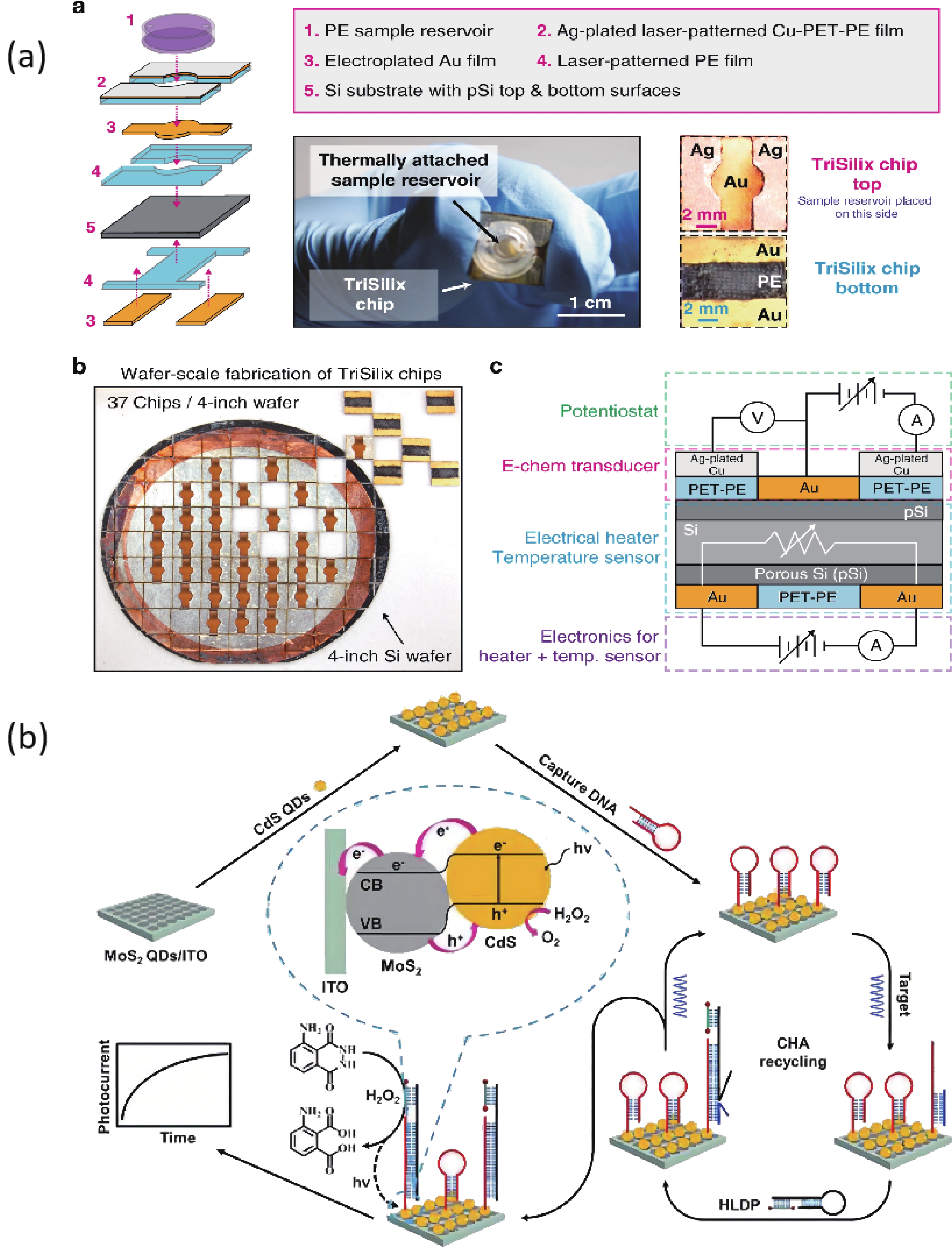
Si is the most commonly used material in large-scale semiconductor biosensors manufacturing, which can greatly reduce the fabricating costs. A portable and battery-powered PCR device which integrated a PCR thermal control system, a PCR reaction chip and the end-point fluorescence detection system was developed by Jie et al. The disposable reaction chips were fabricated with a silicon substrate due to its excellent thermal conductivity, silicone rubber and a quartz plate. The PCR thermal control system was developed using Al thin-film heaters and RTDs to provide fast and precise temperature conditions with an average heating rate of 32 °C/s and an average cooling rate of 7.5 °C/s. The time consumption of the 40 cycles of PCR was less than 1 h[47]. Sun et al. realized to detect five equine respiratory diseases pathogens simultaneously on a silicon microfluidic chip using digital LAMP. Silicon chips are highly stable, have no auto-fluorescence and can be manufactured easily and efficiently. The chips contained ten parallel flow channels with a volume of 1 μL each. The chip surface was thermally oxidized to grow a 200-nm layer of SiO2 that used to reduce the potential for bare silicon to inhibit the amplification process. The detection took place within 30 min with LOD of 18 copies per reaction[48]. Powell and colleagues developed rapid and sensitive nucleic acid amplification tests for HCV, HIV, Zika, HPV16 and HPV18 on the same silicon microchip platform integrated nucleic acid extraction and PCR. These on-chip assays were performed in under 25 min and the efficiency was similar to the assay performed on a bench scale real-time PCR instrument[49]. Nunez-Bajo and co-workers reported a disposable, ultra-low-cost integrated silicon-based on-site transducer (TriSilix) for pathogen nucleic acid amplification and real-time detection. Si was selected as the substrate material to make itself can be used both as an electrical heater and temperature sensor without adding any additional components for temperature transduction. Both isothermal and cyclic nucleic acid amplification can be performed and it integrated temperature control and DNA detection on the same device, which was label-free, requires minimal sample handling. And the TriSilix chips did not need cleanroom for fabrication and can be produced in a standard wet-lab with easily accessible laboratory equipment worldwide. TriSilix was an ideally low-cost, high-performance, on-site nucleic acid detection technology[50] (Fig. 2).

ITO is a conductive and transparent oxide that has advantages of high stability, ease of combination with microfabrication processes, which is used as an electrode material for biosensors[51]. Barreda-Garcia and co-workers utilized the advantages of ITO to develop an optimized and nucleic acid amplification-based biosensor for detecting salmonella. They designed an electrochemical DNA sensor based on a sandwich hybridization assay on the surface of ITO electrodes, which realizes monitoring both PCR and HDA amplicons. ITO was used as a thin film heating element and the reaction surface[52, 53].

Molybdenum disulfide (MoS2) is a typical transition-metal dichalcogenide semiconductor which has unique optical, electronic and catalytic properties. Zang and coauthors developed a CdS/MoS2 heterojunction-based photoelectrochemical DNA biosensor. The CdS/MoS2 photocathode was produced by the stepwise assembly of MoS2 and CdS QDs on ITO. The dual hemin-labeled DNA probe was introduced on high-efficiency CdS/MoS2 electrode via catalytic hairpin assembly (CHA) cycling reaction. This ultrasensitive biosensor achieved a detection limit of 0.39 fM[54] (Fig. 2).

Fig. 2.  (Color online) (a) Schematic illustration of a TriSilix chip and photographs of the actual device[50]. Reproduced with permission. Copyright 2020, Springer Nature. (b) Schematic illustration for CdS/MoS2 heterojunction-enhanced photoelectrochemical sensing strategy coupling with CHA and hemin-mediated chemiluminescence[54]. Reproduced with permission. Copyright 2016, Elsevier.

The principle of nucleic acid amplification-free biosensors depends on the hybridization through spontaneous hydrogen bonding between the target nucleic acid and its complementary strand[55]. The biosensors are usually utilized by immobilizing the single-stranded DNA onto a suitable surface. Au nanorods, CdTe QDs and metallic oxide and so on are growing frequently used to fabricate sensor surface as a promising semiconductor material.

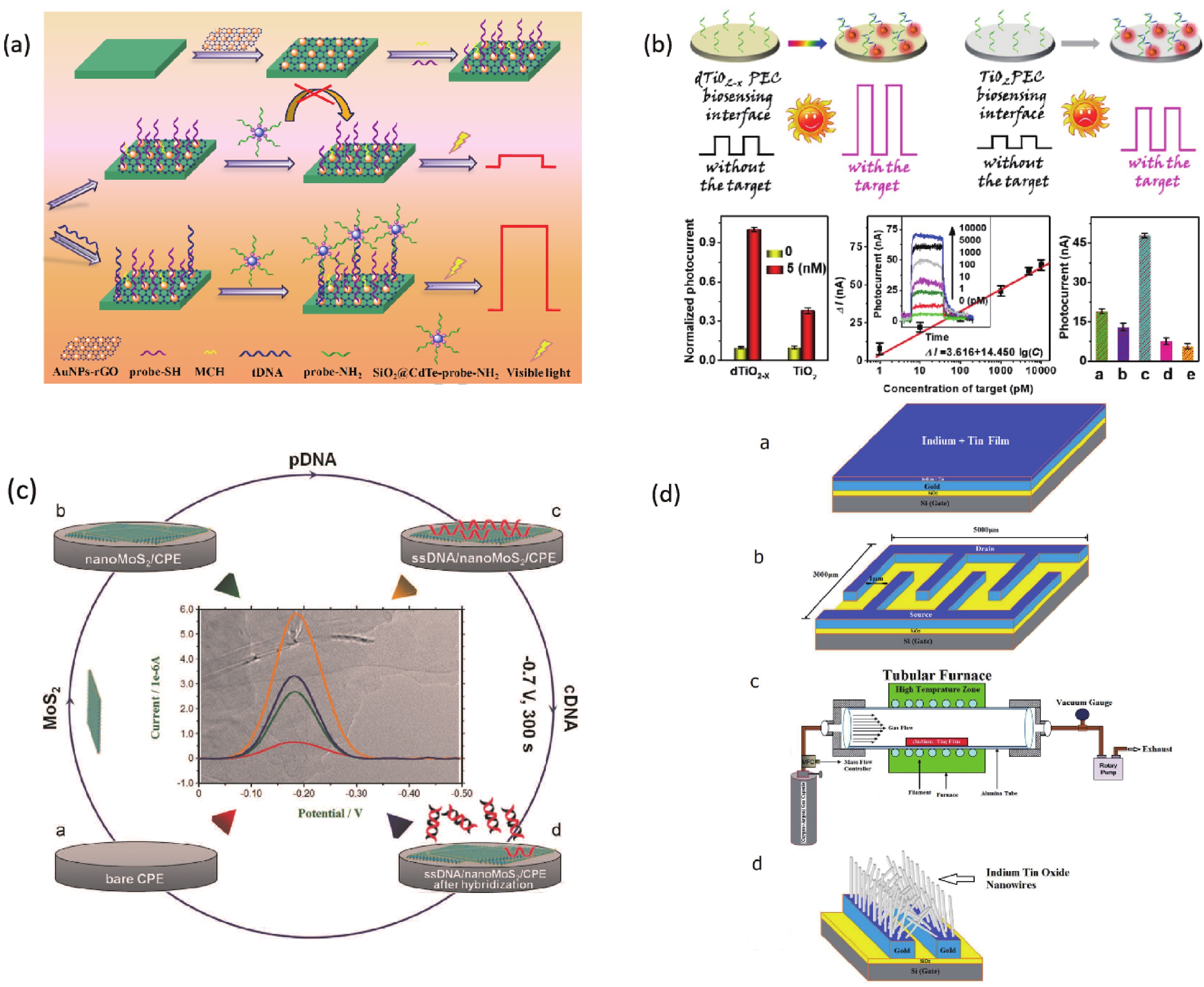
A biosensor for the Cauliflower Mosaic Virus 35S (CaMV35S) promoter detection was reported to distinguish transgenic soybean from non-transgenic samples. The Au nanoparticles-reduced graphene oxide acted as a nanocarrier to immobilize the thiol-functional probe, and the SiO2@CdTe QDs core-shell nanoparticles tagged with the amino-functional probe acted as signal indicators, respectively. In the presence of target DNA of CaMV35S, the binding of tDNA with two probes through the high specific DNA hybridization led to the fabrication of sandwich structure, and thus the high loading of the signal indicators SiO2@CdTe QDs at the electrode surface, which increased the PEC signal. The increased signal depended on the concentration of tDNA and the low detection limit of 0.05 pM was obtained[56] (Fig. 3). Zhang et al. found that monolayer MoS2 has high fluorescence quenching efficiency and shows different affinity towards the single-stranded DNA and double stranded DNA. Therefore, a thin-layer MoS2 nanosheets based and label-free DNA sensor was constructed, with a detection limit of 1.9 × 10⁻¹⁷ M[58, 59] (Fig. 3).

TiO2 is a classical n-type semiconductor, its production method is simple. The raw materials are cheap and easily available. More importantly, it has good biocompatibility, excellent photostability, fast charge conduction rate and excellent photoelectric chemical activity. Many biosensors have been achieved high-performance based on TiO2[60]. A photoelectrochemical DNA sensor based on FTO/TiO2/Au electrode was successfully developed by Liu and coworkers. Combining the small band gap of CdSe QDs with the large band gap of TiO2, the photoelectric conversion efficiency was enhanced. The limit of detection was 3 fM[61]. Tang’s research group modified the electrode surface with the defect-engineered TiO2-x nanorods as photoelectric materials. A photoelectrochemical biosensor platform was constructed for sensitive analysis of target DNA with the surface plasmon resonance enhancement technology of Au nanoparticles. The introduction of Au NPs can not only further enhance the photoelectric signal response but also improve the analytical performance of the sensor on the target DNA. This signaling format combined with exonuclease III-assisted target recycling amplification shows excellent analytical performance[57] (Fig. 3).

Chen and co-workers developed a nanostructured impedance biosensor to detect hepatitis B virus (HBV) DNA. The anodic aluminum oxide membrane having a uniform nano hemisphere array was selected as the substrate of the sensing electrode. A 30-nm gold film was sputtered onto the Al2O3 layer surface as the electrode, followed by electrochemical deposition of Au nanoparticles on each hemisphere surface. Target HBV DNA was captured by hybridization with the specific probe which immobilized on the nanostructured electrode. The LOD was measured to be 111 copy/mL[62]. Zhu et al. developed a DNA sensor to detect HBV with a Cu2O hollow microsphere that can be rapidly synthesized at room temperature. The device has a sensitive detection response and good selectivity for hybridization detection with a detection limit of 1.0 × 10⁻¹⁰ M. This is mainly due to the high specific surface area of Cu2O nanoparticles and the high probe affinity of the particle surface to ssDNA[63]. Shariati et al. reported an ITO nanowires based DNA biosensor compatible with metal-oxide-semiconductor (CMOS) technology to detect HBV. The immobilization of thiolated HBV single-strand DNA is supported with the employment of ITO nanowires reaching the detection limit of 1 fM[64] (Fig. 3). Shariati’s group also developed the HBV detection biosensor whose heterojunction optical electrodes based on WO3/In2O3 nanowires doped with ITO with high sensitivity and stability[65]. Manzano and co-workers reported a label-free user-friendly electrochemical DNA biosensor for hepatitis A virus (HAV). The hybridization reaction occurs on the disposable electrode made of gold functionalized with specific capture probes. The results are monitored through the oxidative peak potential by using cyclic voltammetry[66].

Fig. 3.  (Color online) (a) Schematic illustration of the fabrication procedure of the CaMV35S promoter detection biosensor based on the application of SiO2@CdTe QDs[56]. Reproduced with permission. Copyright 2016, Elsevier. (b) Schematic illustration of biosensor strategies based on the application of dTiO2–x[57]. Reproduced with permission. Copyright 2018, ACS. (c) Schematic illustration of the label-free electrochemical DNA assay based on nanoMoS2 modified electrode (nanoMoS2/CPE)[59]. Reproduced with permission. Copyright 2015, Elsevier. (d) Schematic illustration of the fabrication of ITO nanowires device[64]. Reproduced with permission. Copyright 2018, Elsevier.

Considerable efforts of researches demonstrate the utility of robust nucleic acid tests biosensors with semiconductor for development of a flexible nucleic acid diagnostic platform. With the advantages of semiconductor materials, these biosensors have their own characteristics in rapidity, low cost, high sensitivity and specificity. Accurate and sensitive bioanalysis of nucleic acids is crucial for earlier detection of salmonids viral diseases. Currently, there are no practical applications of semiconductor-based biosensors for salmonids virus detection, but these biosensors have great potential application prospects for the diagnosis of salmonids virus infections in the field. Their portable, rapid and sensitive prosperities could meet the needs of salmonids diseases detection and achieve the goal of early detection of salmonids virus infections.

In spite of wide application prospects of biosensors in salmonids virus detection, there are still some challenges in on-site virus detection and surveillance in salmonids aquaculture. The development trends of on-site virus detection biosensors are mainly in these aspects: (1) Miniaturization and integration of the detection system. It will accord with the diagnostic needs of point-of-care (POC) in resource limited settings such as fish farms. (2) High availability and low cost. Fabricating biosensors can use the lower-cost and easily available semiconductor materials. (3) Multiplexed detection. This is highly desirable in early salmonids virus detection because virus has genetic variability such as IPNV. (4) Application effect evaluation. All biosensors need to be applied in the practical environment to evaluate their detection effect and performance. These goals will encourage researchers to make great progress in more accurate, portable, ultracompact reliable and smart on-site virus detection[67, 68].

The authors thank anonymous reviewers for their constructive comments on the resentation of this article. This research was supported by the National Key Research and Development Program of China (2022YFC2601304) and National Key Research and Development Program of China (2022YFC2602100)


[1]
Bianchi M C, Chopin F, Farme T, et al. FAO: the state of world fisheries and aquaculture. Rome, Italy: Food and Agriculture Organization of the United Nations, 2014
[2]
Crane M, Hyatt A. Viruses of fish: An overview of significant pathogens. Viruses, 2011, 3, 2025 doi: 10.3390/v3112025
[3]
Pinheiro A C A S, Volpe E, Principi D, et al. Development of a multiplex RT-PCR assay for simultaneous detection of the major viruses that affect rainbow trout (Oncorhynchus mykiss). Aquacult Int, 2016, 24, 115 doi: 10.1007/s10499-015-9912-9
[4]
Rodriguez Saint-Jean S, Borrego J J, Perez-Prieto S I. Comparative evaluation of five serological methods and RT-PCR assay for the detection of IPNV in fish. J Virol Methods, 2001, 97, 23 doi: 10.1016/S0166-0934(01)00329-9
[5]
Hoferer M, Akimkin V, Skrypski J, et al. Improvement of a diagnostic procedure in surveillance of the listed fish diseases IHN and VHS. J Fish Dis, 2019, 42, 559 doi: 10.1111/jfd.12968
[6]
Shao Y Z, Zhao J Z, Ren G M, et al. Early or simultaneous infection with infectious pancreatic necrosis virus inhibits infectious hematopoietic necrosis virus replication and induces a stronger antiviral response during Co-infection in rainbow trout (Oncorhynchus mykiss). Viruses, 2022, 14, 1732 doi: 10.3390/v14081732
[7]
Menon S, Mathew M R, Sam S, et al. Recent advances and challenges in electrochemical biosensors for emerging and re-emerging infectious diseases. J Electroanal Chem, 2020, 878, 114596 doi: 10.1016/j.jelechem.2020.114596
[8]
Bhalla N, Jolly P, Formisano N, et al. Introduction to biosensors. Essays Biochem, 2016, 60, 1 doi: 10.1042/EBC20150001
[9]
Kim U, Ghanbari S, Ravikumar A, et al. Rapid, affordable, and point-of-care water monitoring via a microfluidic DNA sensor and a mobile interface for global health. IEEE J Transl Eng Health Med, 2013, 1, 3700207 doi: 10.1109/JTEHM.2013.2281819
[10]
Gavrilescu M, Demnerová K, Aamand J, et al. Emerging pollutants in the environment: Present and future challenges in biomonitoring, ecological risks and bioremediation. New Biotechnol, 2015, 32, 147 doi: 10.1016/j.nbt.2014.01.001
[11]
Chen Y, Wang Z Z, Liu Y X, et al. Recent advances in rapid pathogen detection method based on biosensors. Eur J Clin Microbiol Infect Dis, 2018, 37, 1021 doi: 10.1007/s10096-018-3230-x
[12]
Pandey C M, Augustine S, Kumar S, et al. Microfluidics based point-of-care diagnostics. Biotechnol J, 2018, 13, 1700047 doi: 10.1002/biot.201700047
[13]
Amar E C, Kiron V, Akutsu T, et al. Resistance of rainbow trout Oncorhynchus mykiss to infectious hematopoietic necrosis virus (IHNV) experimental infection following ingestion of natural and synthetic carotenoids. Aquaculture, 2012, 330, 148 doi: 10.1016/j.aquaculture.2011.12.007
[14]
Ahmadivand S, Soltani M, Mardani K, et al. Infectious hematopoietic necrosis virus (IHNV) outbreak in farmed rainbow trout in Iran: Viral isolation, pathological findings, molecular confirmation, and genetic analysis. Virus Res, 2017, 229, 17 doi: 10.1016/j.virusres.2016.12.013
[15]
WOAH. Infection with infectious haematopoietic necrosis virus. Manual of Diagnostic Tests for Aquatic Animals, 2021, chapter 2.3.5
[16]
Ziafati Kafi Z, Ghalyanchilangeroudi A, Nikaein D, et al. Phylogenetic analysis and genotyping of Iranian infectious haematopoietic necrosis virus (IHNV) of rainbow trout (Oncorhynchus mykiss) based on the glycoprotein gene. Vet Med Sci, 2022, 8, 2411 doi: 10.1002/vms3.931
[17]
Kurath G, Winton J R, Dale O B, et al. Atlantic salmon, Salmo salar L. are broadly susceptible to isolates representing the North American genogroups of infectious hematopoietic necrosis virus. J Fish Dis, 2016, 39, 55 doi: 10.1111/jfd.12323
[18]
Tapia D, Eissler Y, Torres P, et al. Detection and phylogenetic analysis of infectious pancreatic necrosis virus in Chile. Dis Aquat Organ, 2015, 116, 173 doi: 10.3354/dao02912
[19]
Zhu L, Wang X L, Wang K Y, et al. Outbreak of infectious pancreatic necrosis virus (IPNV) in farmed rainbow trout in China. Acta Trop, 2017, 170, 63 doi: 10.1016/j.actatropica.2017.02.025
[20]
Suebsing R, Oh M J, Kim J H. Evaluation of rapid and sensitive reverse transcription loop-mediated isothermal amplification method for detecting infectious pancreatic necrosis virus in chum salmon (Oncorhynchus Keta). J Vet Diagn Invest, 2011, 23, 704 doi: 10.1177/1040638711407897
[21]
Yong C Y, Ong H K, Tang H C, et al. Infectious hematopoietic necrosis virus: Advances in diagnosis and vaccine development. PeerJ, 2019, 7, e7151 doi: 10.7717/peerj.7151
[22]
Ning B, Huang Z, Youngquist B M, et al. Liposome-mediated detection of SARS-CoV-2 RNA-positive extracellular vesicles in plasma. Nat Nanotechnol, 2021, 16, 1039 doi: 10.1038/s41565-021-00939-8
[23]
Cella L N, Blackstock D, Yates M A, et al. Detection of RNA viruses: Current technologies and future perspectives. Crit Rev Eukaryot Gene Expr, 2013, 23, 125 doi: 10.1615/CritRevEukaryotGeneExpr.2013006974
[24]
Dhar A K, Bowers R M, Licon K S, et al. Detection and quantification of infectious hematopoietic necrosis virus in rainbow trout (Oncorhynchus mykiss) by SYBR Green real-time reverse transcriptase-polymerase chain reaction. J Virol Methods, 2008, 147, 157 doi: 10.1016/j.jviromet.2007.08.026
[25]
Liu Z X, Teng Y, Liu H, et al. Simultaneous detection of three fish rhabdoviruses using multiplex real-time quantitative RT-PCR assay. J Virol Methods, 2008, 149, 103 doi: 10.1016/j.jviromet.2007.12.017
[26]
Purcell M K, Thompson R L, Garver K A, et al. Universal reverse-transcriptase real-time PCR for infectious hematopoietic necrosis virus (IHNV). Dis Aquat Organ, 2013, 106, 103 doi: 10.3354/dao02644
[27]
Kerr C C, Cunningham C O. Moving molecular diagnostics from laboratory to clinical application: A case study using infectious pancreatic necrosis virus serotype A. Lett Appl Microbiol, 2006, 43, 98 doi: 10.1111/j.1472-765X.2006.01885.x
[28]
Alonso M, Rodríguez S, Pérez-Prieto S I. Viral coinfection in salmonids: Infectious pancreatic necrosis virus interferes with infectious hematopoietic necrosis virus. Arch Virol, 1999, 144, 657 doi: 10.1007/s007050050534
[29]
Hoferer M, Braun A, Skrypski J, et al. One-step cross-genogroup multiplex RT-qPCR with an internal control system for the detection of infectious pancreatic necrosis virus (IPNV). J Virol Methods, 2017, 247, 68 doi: 10.1016/j.jviromet.2017.05.015
[30]
Orpetveit I, Mikalsen A B, Sindre H, et al. Detection of infectious pancreatic necrosis virus in subclinically infected Atlantic salmon by virus isolation in cell culture or real-time reverse transcription polymerase chain reaction: Influence of sample preservation and storage. J Vet Diagn Invest, 2010, 22, 886 doi: 10.1177/104063871002200606
[31]
Bowers R M, Lapatra S E, Dhar A K. Detection and quantitation of infectious pancreatic necrosis virus by real-time reverse transcriptase-polymerase chain reaction using lethal and non-lethal tissue sampling. J Virol Methods, 2008, 147, 226 doi: 10.1016/j.jviromet.2007.09.003
[32]
Jia P, Purcell M K, Pan G, et al. Analytical validation of a reverse transcriptase droplet digital PCR (RT-ddPCR) for quantitative detection of infectious hematopoietic necrosis virus. J Virol Methods, 2017, 245, 73 doi: 10.1016/j.jviromet.2017.03.010
[33]
Cuenca A, Vendramin N, Olesen N J. Analytical validation of one-step realtime RT-PCR for detection of infectious hematopoietic necrosis virus (IHNV). Bull Eur Ass Fish Pathol, 2020, 40, 261
[34]
Suebsing R, Kim J H, Kim S R, et al. Detection of viruses in farmed rainbow trout (Oncorhynchus mykiss) in Korea by RT-LAMP assay. J Microbiol, 2011, 49, 741 doi: 10.1007/s12275-011-1209-8
[35]
Suebsing R, Jeon C H, Oh M J, et al. Reverse transcriptase loop-mediated isothermal amplification assay for infectious hematopoietic necrosis virus in Oncorhynchus Keta. Dis Aquat Organ, 2011, 94, 1 doi: 10.3354/dao02310
[36]
Gunimaladevi I, Kono T, Lapatra S E, et al. A loop mediated isothermal amplification (LAMP) method for detection of infectious hematopoietic necrosis virus (IHNV) in rainbow trout (Oncorhynchus mykiss). Arch Virol, 2005, 150, 899 doi: 10.1007/s00705-004-0468-7
[37]
Soliman H, Midtlyng P J, El-Matbouli M. Sensitive and rapid detection of infectious pancreatic necrosis virus by reverse transcription loop mediated isothermal amplification. J Virol Methods, 2009, 158, 77 doi: 10.1016/j.jviromet.2009.01.018
[38]
Zambry N S, Obande G A, Khalid M F, et al. Utilizing electrochemical-based sensing approaches for the detection of SARS-CoV-2 in clinical samples: A review. Biosensors, 2022, 12, 473 doi: 10.3390/bios12070473
[39]
Chen H, Liu K K, Li Z, et al. Point of care testing for infectious diseases. Clin Chimica Acta, 2019, 493, 138 doi: 10.1016/j.cca.2019.03.008
[40]
Zang Y, Fan J, Ju Y, et al. Current advances in semiconductor nanomaterial-based photoelectrochemical biosensing. Chemistry, 2018, 24, 14010 doi: 10.1002/chem.201801358
[41]
Tay A, Pavesi A, Yazdi S R, et al. Advances in microfluidics in combating infectious diseases. Biotechnol Adv, 2016, 34, 404 doi: 10.1016/j.biotechadv.2016.02.002
[42]
Escobar A, Chiu P, Qu J X, et al. Integrated microfluidic-based platforms for on-site detection and quantification of infectious pathogens: Towards on-site medical translation of SARS-CoV-2 diagnostic platforms. Micromachines, 2021, 12, 1079 doi: 10.3390/mi12091079
[43]
Zhang L, Ding B Z, Chen Q H, et al. Point-of-care-testing of nucleic acids by microfluidics. Trac Trends Anal Chem, 2017, 94, 106 doi: 10.1016/j.trac.2017.07.013
[44]
Jagannath A, Cong H J, Hassan J, et al. Pathogen detection on microfluidic platforms: Recent advances, challenges, and prospects. Biosens Bioelectron X, 2022, 10, 100134 doi: 10.1016/j.biosx.2022.100134
[45]
Wu Q Y, Zhang Y Z, Yang Q, et al. Review of electrochemical DNA biosensors for detecting food borne pathogens. Sensors, 2019, 19, 4916 doi: 10.3390/s19224916
[46]
Maffert P, Reverchon S, Nasser W, et al. New nucleic acid testing devices to diagnose infectious diseases in resource-limited settings. Eur J Clin Microbiol Infect Dis, 2017, 36, 1717 doi: 10.1007/s10096-017-3013-9
[47]
Jie J Y, Hu S M, Liu W W, et al. Portable and battery-powered PCR device for DNA amplification and fluorescence detection. Sensors, 2020, 20, 2627 doi: 10.3390/s20092627
[48]
Sun F, Ganguli A, Nguyen J, et al. Smartphone-based multiplex 30-minute nucleic acid test of live virus from nasal swab extract. Lab Chip, 2020, 20, 1621 doi: 10.1039/D0LC00304B
[49]
Powell L, Wiederkehr R S, Damascus P, et al. Rapid and sensitive detection of viral nucleic acids using silicon microchips. Analyst, 2018, 143, 2596 doi: 10.1039/C8AN00552D
[50]
Nunez-Bajo E, Silva Pinto Collins A, Kasimatis M, et al. Disposable silicon-based all-in-one micro-qPCR for rapid on-site detection of pathogens. Nat Commun, 2020, 11, 6176 doi: 10.1038/s41467-020-19911-6
[51]
Blair E O, Corrigan D K. A review of microfabricated electrochemical biosensors for DNA detection. Biosens Bioelectron, 2019, 134, 57 doi: 10.1016/j.bios.2019.03.055
[52]
Barreda-García S, Miranda-Castro R, de-Los-Santos-Álvarez N, et al. Solid-phase helicase dependent amplification and electrochemical detection of Salmonella on highly stable oligonucleotide-modified ITO electrodes. Chem Commun, 2017, 53, 9721 doi: 10.1039/C7CC05128J
[53]
Barreda-García S, Miranda-Castro R, de-los-Santos-Álvarez N, et al. Sequence-specific electrochemical detection of enzymatic amplification products of Salmonella genome on ITO electrodes improves pathogen detection to the single copy level. Sens Actuat B, 2018, 268, 438 doi: 10.1016/j.snb.2018.04.133
[54]
Zang Y, Lei J P, Hao Q, et al. CdS/MoS2 heterojunction-based photoelectrochemical DNA biosensor via enhanced chemiluminescence excitation. Biosens Bioelectron, 2016, 77, 557 doi: 10.1016/j.bios.2015.10.010
[55]
Asal M, Özen Ö, Şahinler M, et al. Recent developments in enzyme, DNA and immuno-based biosensors. Sensors, 2018, 18, 1924 doi: 10.3390/s18061924
[56]
Li Y Q, Sun L, Liu Q, et al. Photoelectrochemical CaMV35S biosensor for discriminating transgenic from non-transgenic soybean based on SiO2@CdTe quantum dots core-shell nanoparticles as signal indicators. Talanta, 2016, 161, 211 doi: 10.1016/j.talanta.2016.08.047
[57]
Shu J, Qiu Z L, Lv S Z, et al. Plasmonic enhancement coupling with defect-engineered TiO2–x: A mode for sensitive photoelectrochemical biosensing. Anal Chem, 2018, 90, 2425 doi: 10.1021/acs.analchem.7b05296
[58]
Zhu C F, Zeng Z Y, Li H, et al. Single-layer MoS2-based nanoprobes for homogeneous detection of biomolecules. J Am Chem Soc, 2013, 135, 5998 doi: 10.1021/ja4019572
[59]
Wang X X, Nan F X, Zhao J L, et al. A label-free ultrasensitive electrochemical DNA sensor based on thin-layer MoS2 nanosheets with high electrochemical activity. Biosens Bioelectron, 2015, 64, 386 doi: 10.1016/j.bios.2014.09.030
[60]
Ma X G, Wang C, Wu F X, et al. TiO2 nanomaterials in photoelectrochemical and electrochemiluminescent biosensing. Top Curr Chem, 2020, 378, 28 doi: 10.1007/s41061-020-0291-y
[61]
Liu X P, Chen J S, Mao C J, et al. Enhanced photoelectrochemical DNA sensor based on TiO2/Au hybrid structure. Biosens Bioelectron, 2018, 116, 23 doi: 10.1016/j.bios.2018.05.036
[62]
Chen C C, Lai Z L, Wang G J, et al. Polymerase chain reaction-free detection of hepatitis B virus DNA using a nanostructured impedance biosensor. Biosens Bioelectron, 2016, 77, 603 doi: 10.1016/j.bios.2015.10.028
[63]
Zhu H T, Wang J X, Xu G Y. Fast synthesis of Cu2O hollow microspheres and their application in DNA biosensor of hepatitis B virus. Cryst Growth Des, 2009, 9, 633 doi: 10.1021/cg801006g
[64]
Shariati M. The field effect transistor DNA biosensor based on ITO nanowires in label-free hepatitis B virus detecting compatible with CMOS technology. Biosens Bioelectron, 2018, 105, 58 doi: 10.1016/j.bios.2018.01.022
[65]
Shariati M, Sadeghi M. Ultrasensitive DNA biosensor for hepatitis B virus detection based on tin-doped WO3/In2O3 heterojunction nanowire photoelectrode under laser amplification. Anal Bioanal Chem, 2020, 412, 5367 doi: 10.1007/s00216-020-02752-z
[66]
Manzano M, Viezzi S, Mazerat S, et al. Rapid and label-free electrochemical DNA biosensor for detecting hepatitis A virus. Biosens Bioelectron, 2018, 100, 89 doi: 10.1016/j.bios.2017.08.043
[67]
Lim R R X, Bonanni A. The potential of electrochemistry for the detection of coronavirus-induced infections. Trac Trends Anal Chem, 2020, 133, 116081 doi: 10.1016/j.trac.2020.116081
[68]
Pumford E A, Lu J K, Spaczai I, et al. Developments in integrating nucleic acid isothermal amplification and detection systems for point-of-care diagnostics. Biosens Bioelectron, 2020, 170, 112674 doi: 10.1016/j.bios.2020.112674
Fig. 1.  (Color online) The traditional workflow of salmonids diseases detection and surveillance.

Fig. 2.  (Color online) (a) Schematic illustration of a TriSilix chip and photographs of the actual device[50]. Reproduced with permission. Copyright 2020, Springer Nature. (b) Schematic illustration for CdS/MoS2 heterojunction-enhanced photoelectrochemical sensing strategy coupling with CHA and hemin-mediated chemiluminescence[54]. Reproduced with permission. Copyright 2016, Elsevier.

Fig. 3.  (Color online) (a) Schematic illustration of the fabrication procedure of the CaMV35S promoter detection biosensor based on the application of SiO2@CdTe QDs[56]. Reproduced with permission. Copyright 2016, Elsevier. (b) Schematic illustration of biosensor strategies based on the application of dTiO2–x[57]. Reproduced with permission. Copyright 2018, ACS. (c) Schematic illustration of the label-free electrochemical DNA assay based on nanoMoS2 modified electrode (nanoMoS2/CPE)[59]. Reproduced with permission. Copyright 2015, Elsevier. (d) Schematic illustration of the fabrication of ITO nanowires device[64]. Reproduced with permission. Copyright 2018, Elsevier.

[1]
Bianchi M C, Chopin F, Farme T, et al. FAO: the state of world fisheries and aquaculture. Rome, Italy: Food and Agriculture Organization of the United Nations, 2014
[2]
Crane M, Hyatt A. Viruses of fish: An overview of significant pathogens. Viruses, 2011, 3, 2025 doi: 10.3390/v3112025
[3]
Pinheiro A C A S, Volpe E, Principi D, et al. Development of a multiplex RT-PCR assay for simultaneous detection of the major viruses that affect rainbow trout (Oncorhynchus mykiss). Aquacult Int, 2016, 24, 115 doi: 10.1007/s10499-015-9912-9
[4]
Rodriguez Saint-Jean S, Borrego J J, Perez-Prieto S I. Comparative evaluation of five serological methods and RT-PCR assay for the detection of IPNV in fish. J Virol Methods, 2001, 97, 23 doi: 10.1016/S0166-0934(01)00329-9
[5]
Hoferer M, Akimkin V, Skrypski J, et al. Improvement of a diagnostic procedure in surveillance of the listed fish diseases IHN and VHS. J Fish Dis, 2019, 42, 559 doi: 10.1111/jfd.12968
[6]
Shao Y Z, Zhao J Z, Ren G M, et al. Early or simultaneous infection with infectious pancreatic necrosis virus inhibits infectious hematopoietic necrosis virus replication and induces a stronger antiviral response during Co-infection in rainbow trout (Oncorhynchus mykiss). Viruses, 2022, 14, 1732 doi: 10.3390/v14081732
[7]
Menon S, Mathew M R, Sam S, et al. Recent advances and challenges in electrochemical biosensors for emerging and re-emerging infectious diseases. J Electroanal Chem, 2020, 878, 114596 doi: 10.1016/j.jelechem.2020.114596
[8]
Bhalla N, Jolly P, Formisano N, et al. Introduction to biosensors. Essays Biochem, 2016, 60, 1 doi: 10.1042/EBC20150001
[9]
Kim U, Ghanbari S, Ravikumar A, et al. Rapid, affordable, and point-of-care water monitoring via a microfluidic DNA sensor and a mobile interface for global health. IEEE J Transl Eng Health Med, 2013, 1, 3700207 doi: 10.1109/JTEHM.2013.2281819
[10]
Gavrilescu M, Demnerová K, Aamand J, et al. Emerging pollutants in the environment: Present and future challenges in biomonitoring, ecological risks and bioremediation. New Biotechnol, 2015, 32, 147 doi: 10.1016/j.nbt.2014.01.001
[11]
Chen Y, Wang Z Z, Liu Y X, et al. Recent advances in rapid pathogen detection method based on biosensors. Eur J Clin Microbiol Infect Dis, 2018, 37, 1021 doi: 10.1007/s10096-018-3230-x
[12]
Pandey C M, Augustine S, Kumar S, et al. Microfluidics based point-of-care diagnostics. Biotechnol J, 2018, 13, 1700047 doi: 10.1002/biot.201700047
[13]
Amar E C, Kiron V, Akutsu T, et al. Resistance of rainbow trout Oncorhynchus mykiss to infectious hematopoietic necrosis virus (IHNV) experimental infection following ingestion of natural and synthetic carotenoids. Aquaculture, 2012, 330, 148 doi: 10.1016/j.aquaculture.2011.12.007
[14]
Ahmadivand S, Soltani M, Mardani K, et al. Infectious hematopoietic necrosis virus (IHNV) outbreak in farmed rainbow trout in Iran: Viral isolation, pathological findings, molecular confirmation, and genetic analysis. Virus Res, 2017, 229, 17 doi: 10.1016/j.virusres.2016.12.013
[15]
WOAH. Infection with infectious haematopoietic necrosis virus. Manual of Diagnostic Tests for Aquatic Animals, 2021, chapter 2.3.5
[16]
Ziafati Kafi Z, Ghalyanchilangeroudi A, Nikaein D, et al. Phylogenetic analysis and genotyping of Iranian infectious haematopoietic necrosis virus (IHNV) of rainbow trout (Oncorhynchus mykiss) based on the glycoprotein gene. Vet Med Sci, 2022, 8, 2411 doi: 10.1002/vms3.931
[17]
Kurath G, Winton J R, Dale O B, et al. Atlantic salmon, Salmo salar L. are broadly susceptible to isolates representing the North American genogroups of infectious hematopoietic necrosis virus. J Fish Dis, 2016, 39, 55 doi: 10.1111/jfd.12323
[18]
Tapia D, Eissler Y, Torres P, et al. Detection and phylogenetic analysis of infectious pancreatic necrosis virus in Chile. Dis Aquat Organ, 2015, 116, 173 doi: 10.3354/dao02912
[19]
Zhu L, Wang X L, Wang K Y, et al. Outbreak of infectious pancreatic necrosis virus (IPNV) in farmed rainbow trout in China. Acta Trop, 2017, 170, 63 doi: 10.1016/j.actatropica.2017.02.025
[20]
Suebsing R, Oh M J, Kim J H. Evaluation of rapid and sensitive reverse transcription loop-mediated isothermal amplification method for detecting infectious pancreatic necrosis virus in chum salmon (Oncorhynchus Keta). J Vet Diagn Invest, 2011, 23, 704 doi: 10.1177/1040638711407897
[21]
Yong C Y, Ong H K, Tang H C, et al. Infectious hematopoietic necrosis virus: Advances in diagnosis and vaccine development. PeerJ, 2019, 7, e7151 doi: 10.7717/peerj.7151
[22]
Ning B, Huang Z, Youngquist B M, et al. Liposome-mediated detection of SARS-CoV-2 RNA-positive extracellular vesicles in plasma. Nat Nanotechnol, 2021, 16, 1039 doi: 10.1038/s41565-021-00939-8
[23]
Cella L N, Blackstock D, Yates M A, et al. Detection of RNA viruses: Current technologies and future perspectives. Crit Rev Eukaryot Gene Expr, 2013, 23, 125 doi: 10.1615/CritRevEukaryotGeneExpr.2013006974
[24]
Dhar A K, Bowers R M, Licon K S, et al. Detection and quantification of infectious hematopoietic necrosis virus in rainbow trout (Oncorhynchus mykiss) by SYBR Green real-time reverse transcriptase-polymerase chain reaction. J Virol Methods, 2008, 147, 157 doi: 10.1016/j.jviromet.2007.08.026
[25]
Liu Z X, Teng Y, Liu H, et al. Simultaneous detection of three fish rhabdoviruses using multiplex real-time quantitative RT-PCR assay. J Virol Methods, 2008, 149, 103 doi: 10.1016/j.jviromet.2007.12.017
[26]
Purcell M K, Thompson R L, Garver K A, et al. Universal reverse-transcriptase real-time PCR for infectious hematopoietic necrosis virus (IHNV). Dis Aquat Organ, 2013, 106, 103 doi: 10.3354/dao02644
[27]
Kerr C C, Cunningham C O. Moving molecular diagnostics from laboratory to clinical application: A case study using infectious pancreatic necrosis virus serotype A. Lett Appl Microbiol, 2006, 43, 98 doi: 10.1111/j.1472-765X.2006.01885.x
[28]
Alonso M, Rodríguez S, Pérez-Prieto S I. Viral coinfection in salmonids: Infectious pancreatic necrosis virus interferes with infectious hematopoietic necrosis virus. Arch Virol, 1999, 144, 657 doi: 10.1007/s007050050534
[29]
Hoferer M, Braun A, Skrypski J, et al. One-step cross-genogroup multiplex RT-qPCR with an internal control system for the detection of infectious pancreatic necrosis virus (IPNV). J Virol Methods, 2017, 247, 68 doi: 10.1016/j.jviromet.2017.05.015
[30]
Orpetveit I, Mikalsen A B, Sindre H, et al. Detection of infectious pancreatic necrosis virus in subclinically infected Atlantic salmon by virus isolation in cell culture or real-time reverse transcription polymerase chain reaction: Influence of sample preservation and storage. J Vet Diagn Invest, 2010, 22, 886 doi: 10.1177/104063871002200606
[31]
Bowers R M, Lapatra S E, Dhar A K. Detection and quantitation of infectious pancreatic necrosis virus by real-time reverse transcriptase-polymerase chain reaction using lethal and non-lethal tissue sampling. J Virol Methods, 2008, 147, 226 doi: 10.1016/j.jviromet.2007.09.003
[32]
Jia P, Purcell M K, Pan G, et al. Analytical validation of a reverse transcriptase droplet digital PCR (RT-ddPCR) for quantitative detection of infectious hematopoietic necrosis virus. J Virol Methods, 2017, 245, 73 doi: 10.1016/j.jviromet.2017.03.010
[33]
Cuenca A, Vendramin N, Olesen N J. Analytical validation of one-step realtime RT-PCR for detection of infectious hematopoietic necrosis virus (IHNV). Bull Eur Ass Fish Pathol, 2020, 40, 261
[34]
Suebsing R, Kim J H, Kim S R, et al. Detection of viruses in farmed rainbow trout (Oncorhynchus mykiss) in Korea by RT-LAMP assay. J Microbiol, 2011, 49, 741 doi: 10.1007/s12275-011-1209-8
[35]
Suebsing R, Jeon C H, Oh M J, et al. Reverse transcriptase loop-mediated isothermal amplification assay for infectious hematopoietic necrosis virus in Oncorhynchus Keta. Dis Aquat Organ, 2011, 94, 1 doi: 10.3354/dao02310
[36]
Gunimaladevi I, Kono T, Lapatra S E, et al. A loop mediated isothermal amplification (LAMP) method for detection of infectious hematopoietic necrosis virus (IHNV) in rainbow trout (Oncorhynchus mykiss). Arch Virol, 2005, 150, 899 doi: 10.1007/s00705-004-0468-7
[37]
Soliman H, Midtlyng P J, El-Matbouli M. Sensitive and rapid detection of infectious pancreatic necrosis virus by reverse transcription loop mediated isothermal amplification. J Virol Methods, 2009, 158, 77 doi: 10.1016/j.jviromet.2009.01.018
[38]
Zambry N S, Obande G A, Khalid M F, et al. Utilizing electrochemical-based sensing approaches for the detection of SARS-CoV-2 in clinical samples: A review. Biosensors, 2022, 12, 473 doi: 10.3390/bios12070473
[39]
Chen H, Liu K K, Li Z, et al. Point of care testing for infectious diseases. Clin Chimica Acta, 2019, 493, 138 doi: 10.1016/j.cca.2019.03.008
[40]
Zang Y, Fan J, Ju Y, et al. Current advances in semiconductor nanomaterial-based photoelectrochemical biosensing. Chemistry, 2018, 24, 14010 doi: 10.1002/chem.201801358
[41]
Tay A, Pavesi A, Yazdi S R, et al. Advances in microfluidics in combating infectious diseases. Biotechnol Adv, 2016, 34, 404 doi: 10.1016/j.biotechadv.2016.02.002
[42]
Escobar A, Chiu P, Qu J X, et al. Integrated microfluidic-based platforms for on-site detection and quantification of infectious pathogens: Towards on-site medical translation of SARS-CoV-2 diagnostic platforms. Micromachines, 2021, 12, 1079 doi: 10.3390/mi12091079
[43]
Zhang L, Ding B Z, Chen Q H, et al. Point-of-care-testing of nucleic acids by microfluidics. Trac Trends Anal Chem, 2017, 94, 106 doi: 10.1016/j.trac.2017.07.013
[44]
Jagannath A, Cong H J, Hassan J, et al. Pathogen detection on microfluidic platforms: Recent advances, challenges, and prospects. Biosens Bioelectron X, 2022, 10, 100134 doi: 10.1016/j.biosx.2022.100134
[45]
Wu Q Y, Zhang Y Z, Yang Q, et al. Review of electrochemical DNA biosensors for detecting food borne pathogens. Sensors, 2019, 19, 4916 doi: 10.3390/s19224916
[46]
Maffert P, Reverchon S, Nasser W, et al. New nucleic acid testing devices to diagnose infectious diseases in resource-limited settings. Eur J Clin Microbiol Infect Dis, 2017, 36, 1717 doi: 10.1007/s10096-017-3013-9
[47]
Jie J Y, Hu S M, Liu W W, et al. Portable and battery-powered PCR device for DNA amplification and fluorescence detection. Sensors, 2020, 20, 2627 doi: 10.3390/s20092627
[48]
Sun F, Ganguli A, Nguyen J, et al. Smartphone-based multiplex 30-minute nucleic acid test of live virus from nasal swab extract. Lab Chip, 2020, 20, 1621 doi: 10.1039/D0LC00304B
[49]
Powell L, Wiederkehr R S, Damascus P, et al. Rapid and sensitive detection of viral nucleic acids using silicon microchips. Analyst, 2018, 143, 2596 doi: 10.1039/C8AN00552D
[50]
Nunez-Bajo E, Silva Pinto Collins A, Kasimatis M, et al. Disposable silicon-based all-in-one micro-qPCR for rapid on-site detection of pathogens. Nat Commun, 2020, 11, 6176 doi: 10.1038/s41467-020-19911-6
[51]
Blair E O, Corrigan D K. A review of microfabricated electrochemical biosensors for DNA detection. Biosens Bioelectron, 2019, 134, 57 doi: 10.1016/j.bios.2019.03.055
[52]
Barreda-García S, Miranda-Castro R, de-Los-Santos-Álvarez N, et al. Solid-phase helicase dependent amplification and electrochemical detection of Salmonella on highly stable oligonucleotide-modified ITO electrodes. Chem Commun, 2017, 53, 9721 doi: 10.1039/C7CC05128J
[53]
Barreda-García S, Miranda-Castro R, de-los-Santos-Álvarez N, et al. Sequence-specific electrochemical detection of enzymatic amplification products of Salmonella genome on ITO electrodes improves pathogen detection to the single copy level. Sens Actuat B, 2018, 268, 438 doi: 10.1016/j.snb.2018.04.133
[54]
Zang Y, Lei J P, Hao Q, et al. CdS/MoS2 heterojunction-based photoelectrochemical DNA biosensor via enhanced chemiluminescence excitation. Biosens Bioelectron, 2016, 77, 557 doi: 10.1016/j.bios.2015.10.010
[55]
Asal M, Özen Ö, Şahinler M, et al. Recent developments in enzyme, DNA and immuno-based biosensors. Sensors, 2018, 18, 1924 doi: 10.3390/s18061924
[56]
Li Y Q, Sun L, Liu Q, et al. Photoelectrochemical CaMV35S biosensor for discriminating transgenic from non-transgenic soybean based on SiO2@CdTe quantum dots core-shell nanoparticles as signal indicators. Talanta, 2016, 161, 211 doi: 10.1016/j.talanta.2016.08.047
[57]
Shu J, Qiu Z L, Lv S Z, et al. Plasmonic enhancement coupling with defect-engineered TiO2–x: A mode for sensitive photoelectrochemical biosensing. Anal Chem, 2018, 90, 2425 doi: 10.1021/acs.analchem.7b05296
[58]
Zhu C F, Zeng Z Y, Li H, et al. Single-layer MoS2-based nanoprobes for homogeneous detection of biomolecules. J Am Chem Soc, 2013, 135, 5998 doi: 10.1021/ja4019572
[59]
Wang X X, Nan F X, Zhao J L, et al. A label-free ultrasensitive electrochemical DNA sensor based on thin-layer MoS2 nanosheets with high electrochemical activity. Biosens Bioelectron, 2015, 64, 386 doi: 10.1016/j.bios.2014.09.030
[60]
Ma X G, Wang C, Wu F X, et al. TiO2 nanomaterials in photoelectrochemical and electrochemiluminescent biosensing. Top Curr Chem, 2020, 378, 28 doi: 10.1007/s41061-020-0291-y
[61]
Liu X P, Chen J S, Mao C J, et al. Enhanced photoelectrochemical DNA sensor based on TiO2/Au hybrid structure. Biosens Bioelectron, 2018, 116, 23 doi: 10.1016/j.bios.2018.05.036
[62]
Chen C C, Lai Z L, Wang G J, et al. Polymerase chain reaction-free detection of hepatitis B virus DNA using a nanostructured impedance biosensor. Biosens Bioelectron, 2016, 77, 603 doi: 10.1016/j.bios.2015.10.028
[63]
Zhu H T, Wang J X, Xu G Y. Fast synthesis of Cu2O hollow microspheres and their application in DNA biosensor of hepatitis B virus. Cryst Growth Des, 2009, 9, 633 doi: 10.1021/cg801006g
[64]
Shariati M. The field effect transistor DNA biosensor based on ITO nanowires in label-free hepatitis B virus detecting compatible with CMOS technology. Biosens Bioelectron, 2018, 105, 58 doi: 10.1016/j.bios.2018.01.022
[65]
Shariati M, Sadeghi M. Ultrasensitive DNA biosensor for hepatitis B virus detection based on tin-doped WO3/In2O3 heterojunction nanowire photoelectrode under laser amplification. Anal Bioanal Chem, 2020, 412, 5367 doi: 10.1007/s00216-020-02752-z
[66]
Manzano M, Viezzi S, Mazerat S, et al. Rapid and label-free electrochemical DNA biosensor for detecting hepatitis A virus. Biosens Bioelectron, 2018, 100, 89 doi: 10.1016/j.bios.2017.08.043
[67]
Lim R R X, Bonanni A. The potential of electrochemistry for the detection of coronavirus-induced infections. Trac Trends Anal Chem, 2020, 133, 116081 doi: 10.1016/j.trac.2020.116081
[68]
Pumford E A, Lu J K, Spaczai I, et al. Developments in integrating nucleic acid isothermal amplification and detection systems for point-of-care diagnostics. Biosens Bioelectron, 2020, 170, 112674 doi: 10.1016/j.bios.2020.112674
1

A family of flexible two-dimensional semiconductors: MgMX2Y6 (M = Ti/Zr/Hf; X = Si/Ge; Y = S/Se/Te)

Junhui Yuan, Kanhao Xue, Xiangshui Miao, Lei Ye

Journal of Semiconductors, 2023, 44(4): 042101. doi: 10.1088/1674-4926/44/4/042101

2

Application and prospect of semiconductor biosensors in detection of viral zoonoses

Jiahao Zheng, Chunyan Feng, Songyin Qiu, Ke Xu, Caixia Wang, et al.

Journal of Semiconductors, 2023, 44(2): 023102. doi: 10.1088/1674-4926/44/2/023102

3

n-Type acceptor–acceptor polymer semiconductors

Yongqiang Shi, Liming Ding

Journal of Semiconductors, 2021, 42(10): 100202. doi: 10.1088/1674-4926/42/10/100202

4

Giant spin injection into semiconductor and THz pulse emission

Zheng Feng

Journal of Semiconductors, 2019, 40(7): 070201. doi: 10.1088/1674-4926/40/7/070201

5

Hybrid functional microfibers for textile electronics and biosensors

Bichitra Nanda Sahoo, Byungwoo Choi, Jungmok Seo, Taeyoon Lee

Journal of Semiconductors, 2018, 39(1): 011009. doi: 10.1088/1674-4926/39/1/011009

6

Studies of Water V. Five Phonons in Protonic Semiconductor Lattice Model of Pure Liquid Water

Binbin Jie, Chihtang Sah

Journal of Semiconductors, 2017, 38(7): 071001. doi: 10.1088/1674-4926/38/7/071001

7

Detection of lead ions with AlGaAs/InGaAs pseudomorphic high electron mobility transistor

Jiqiang Niu, Yang Zhang, Min Guan, Chengyan Wang, Lijie Cui, et al.

Journal of Semiconductors, 2016, 37(11): 114003. doi: 10.1088/1674-4926/37/11/114003

8

Progress in complementary metal-oxide-semiconductor silicon photonics and optoelectronic integrated circuits

Hongda Chen, Zan Zhang, Beiju Huang, Luhong Mao, Zanyun Zhang, et al.

Journal of Semiconductors, 2015, 36(12): 121001. doi: 10.1088/1674-4926/36/12/121001

9

First-principles calculations of Mg2X (X = Si, Ge, Sn) semiconductors with the calcium fluorite structure

Sandong Guo

Journal of Semiconductors, 2015, 36(5): 053002. doi: 10.1088/1674-4926/36/5/053002

10

Large-signal characterizations of DDR IMPATT devices based on group Ⅲ-Ⅴ semiconductors at millimeter-wave and terahertz frequencies

Aritra Acharyya, Aliva Mallik, Debopriya Banerjee, Suman Ganguli, Arindam Das, et al.

Journal of Semiconductors, 2014, 35(8): 084003. doi: 10.1088/1674-4926/35/8/084003

11

The effects of electron irradiation on the optical properties of the organic semiconductor polypyrrole

J. V. Thombare, M. C. Rath, S. H. Han, V. J. Fulari

Journal of Semiconductors, 2013, 34(9): 093001. doi: 10.1088/1674-4926/34/9/093001

12

A theoretical model of the femtosecond laser ablation of semiconductors considering inverse bremsstrahlung absorption

Lin Xiaohui, Zhang Chibin, Ren Weisong, Jiang Shuyun, Ouyang Quanhui, et al.

Journal of Semiconductors, 2012, 33(4): 046002. doi: 10.1088/1674-4926/33/4/046002

13

Microstructural properties of over-doped GaN-based diluted magnetic semiconductors grown by MOCVD

Tao Zhikuo, Zhang Rong, Xiu Xiangqian, Cui Xugao, Li Li, et al.

Journal of Semiconductors, 2012, 33(7): 073002. doi: 10.1088/1674-4926/33/7/073002

14

Electrical characterization of the organic semiconductor Ag/CuPc/Au Schottky diode

Mutabar Shah, M. H. Sayyad, Kh. S. Karimov

Journal of Semiconductors, 2011, 32(4): 044001. doi: 10.1088/1674-4926/32/4/044001

15

Local charge neutrality condition, Fermi level and majority carrier density of a semiconductor with multiple localized multi-level intrinsic/impurity defects

Ken K. Chin

Journal of Semiconductors, 2011, 32(11): 112001. doi: 10.1088/1674-4926/32/11/112001

16

A signal processing method for the friction-based endpoint detection system of a CMP process

Xu Chi, Guo Dongming, Jin Zhuji, Kang Renke

Journal of Semiconductors, 2010, 31(12): 126002. doi: 10.1088/1674-4926/31/12/126002

17

Synthesis of organic semiconductor hexadecachloro zinc phthalocyanine and its gas sensitivity

Shi Yunbo, Lei Tingping, Li Zan, Xiu Debin, Zhao Wenjie, et al.

Journal of Semiconductors, 2009, 30(3): 034009. doi: 10.1088/1674-4926/30/3/034009

18

Terahertz Semiconductor Quantum Well Devices

Liu H C, Luo H, Ban D, Wachter M, Song C Y, et al.

Chinese Journal of Semiconductors , 2006, 27(4): 627-634.

19

Electronic Structure of Semiconductor Nanocrystals

Li Jingbo, Wang Linwang, Wei Suhuai

Chinese Journal of Semiconductors , 2006, 27(2): 191-196.

20

Passively Mode Locked Diode-End-Pumped Yb∶YAB Laser with High Reflectivity Type Semiconductor Saturable Absorption Mirror

Wang Yonggang, Ma Xiaoyu, Xue Yinghong, Sun Hong,Zhang Zhigang,and Wang Qingyue

Chinese Journal of Semiconductors , 2005, 26(2): 250-253.

1. Huang, Y., Lu, Y., Liu, X. et al. A portable all-in-one microfluidic platform integrated with CRISPR-based extraction-free assay for rapid and on-site detection of monkeypox and lumpy skin disease. Sensors and Actuators B: Chemical, 2025. doi:10.1016/j.snb.2025.137612
2. Zhong, B., Xu, H., Qin, X. et al. A crosstalk-free dual-mode sweat sensing system for naked-eye sweat loss quantification via changes in structural reflectance. Bio-Design and Manufacturing, 2024, 7(4): 428-438. doi:10.1007/s42242-024-00294-2
3. Chen, Y., Gao, S., Xu, H. et al. The development and initial evaluation of a multiplex RT-PCR and high-throughput liquid chip assay for the simultaneous detection of five salmonid virus diseases. Aquaculture Reports, 2024. doi:10.1016/j.aqrep.2024.102163
4. Li, Z., Lin, X., He, D. et al. Preface to Special Issue on Novel Semiconductor-biochemical Sensors. Journal of Semiconductors, 2023, 44(2): 020101. doi:10.1088/1674-4926/44/2/020101
  • Search

    Advanced Search >>

    GET CITATION

    Xiaofei Liu, Songyin Qiu, Haiping Fang, Lin Mei, Hongli Jing, Chunyan Feng, Shaoqiang Wu, Xiangmei Lin. A brief review of novel nucleic acid test biosensors and their application prospects for salmonids viral diseases detection[J]. Journal of Semiconductors, 2023, 44(2): 023103. doi: 10.1088/1674-4926/44/2/023103
    X F Liu, S Y Qiu, H P Fang, L Mei, H L Jing, C Y Feng, S Q Wu, X M Lin. A brief review of novel nucleic acid test biosensors and their application prospects for salmonids viral diseases detection[J]. J. Semicond, 2023, 44(2): 023103. doi: 10.1088/1674-4926/44/2/023103
    shu

    Export: BibTex EndNote

    Article Metrics

    Article views: 1051 Times PDF downloads: 43 Times Cited by: 4 Times

    History

    Received: 18 December 2022 Revised: 18 January 2023 Online: Accepted Manuscript: 03 February 2023Uncorrected proof: 06 February 2023Published: 10 February 2023

    Catalog

      Email This Article

      User name:
      Email:*请输入正确邮箱
      Code:*验证码错误
      Xiaofei Liu, Songyin Qiu, Haiping Fang, Lin Mei, Hongli Jing, Chunyan Feng, Shaoqiang Wu, Xiangmei Lin. A brief review of novel nucleic acid test biosensors and their application prospects for salmonids viral diseases detection[J]. Journal of Semiconductors, 2023, 44(2): 023103. doi: 10.1088/1674-4926/44/2/023103 ****X F Liu, S Y Qiu, H P Fang, L Mei, H L Jing, C Y Feng, S Q Wu, X M Lin. A brief review of novel nucleic acid test biosensors and their application prospects for salmonids viral diseases detection[J]. J. Semicond, 2023, 44(2): 023103. doi: 10.1088/1674-4926/44/2/023103
      Citation:
      Xiaofei Liu, Songyin Qiu, Haiping Fang, Lin Mei, Hongli Jing, Chunyan Feng, Shaoqiang Wu, Xiangmei Lin. A brief review of novel nucleic acid test biosensors and their application prospects for salmonids viral diseases detection[J]. Journal of Semiconductors, 2023, 44(2): 023103. doi: 10.1088/1674-4926/44/2/023103 ****
      X F Liu, S Y Qiu, H P Fang, L Mei, H L Jing, C Y Feng, S Q Wu, X M Lin. A brief review of novel nucleic acid test biosensors and their application prospects for salmonids viral diseases detection[J]. J. Semicond, 2023, 44(2): 023103. doi: 10.1088/1674-4926/44/2/023103

      A brief review of novel nucleic acid test biosensors and their application prospects for salmonids viral diseases detection

      DOI: 10.1088/1674-4926/44/2/023103
      More Information
      • Xiaofei Liu:got her Master's Degree on Veterinary in 2014 at Jilin University. Her research interests include detection and monitoring technologies for animal diseases, animal disease risk analysis and genetically modified animals detection
      • Shaoqiang Wu:got his Doctor's Degree on Veterinary Parasitology in 2003 at China Agricultural University. His research interests include precision detection technology for biosecurity risk, on-site detection techniques for shellfish parasites and aquatic animal diseases
      • Xiangmei Lin:got her Doctor's Degree on Veterinary Pathology in 1998 at Nanjing Agricultural University. Her research interests include detection and monitoring technologies for animal diseases, zoonotic diseases, foreign animal diseases and monitoring technologies of genetically modified animals detection
      • Corresponding author: sqwu@sina.comlinxm@caiq.org.cn
      • Received Date: 2022-12-18
      • Revised Date: 2023-01-18
      • Available Online: 2023-02-03

      Catalog

        /

        DownLoad:  Full-Size Img  PowerPoint
        Return
        Return