Processing math: 100%
J. Semicond. > 2025, Volume 46 > Issue 1 > 011604

REVIEWS

Neurotransmitter-mediated artificial synapses based on organic electrochemical transistors for future biomimic and bioinspired neuromorphic systems

Miao Cheng1, 2, 3, §, Yifan Xie1, §, Jinyao Wang1, §, Qingqing Jin1, Yue Tian1, Changrui Liu1, Jingyun Chu1, Mengmeng Li1, 2, 3, and Ling Li1, 2, 3,

+ Author Affiliations

 Corresponding author: Mengmeng Li, limengmeng@ime.ac.cn; Ling Li, lingli@ime.ac.cn

DOI: 10.1088/1674-4926/24090013CSTR: 32376.14.1674-4926.24090013

PDF

Turn off MathJax

Abstract: Organic electrochemical transistors have emerged as a solution for artificial synapses that mimic the neural functions of the brain structure, holding great potentials to break the bottleneck of von Neumann architectures. However, current artificial synapses rely primarily on electrical signals, and little attention has been paid to the vital role of neurotransmitter-mediated artificial synapses. Dopamine is a key neurotransmitter associated with emotion regulation and cognitive processes that needs to be monitored in real time to advance the development of disease diagnostics and neuroscience. To provide insights into the development of artificial synapses with neurotransmitter involvement, this review proposes three steps towards future biomimic and bioinspired neuromorphic systems. We first summarize OECT-based dopamine detection devices, and then review advances in neurotransmitter-mediated artificial synapses and resultant advanced neuromorphic systems. Finally, by exploring the challenges and opportunities related to such neuromorphic systems, we provide a perspective on the future development of biomimetic and bioinspired neuromorphic systems.

Key words: artificial synapsesorganic electrochemical transistorsneurotransmittersneuromorphic systems

The nervous system of all organisms, including brain structures, is composed of two types of synapses: electrical and chemical. In electrical synapses, the cytoplasm of adjacent neuros is directly connected by gap junctions, allowing the bidirectional passage of electrical currents. In contrast, chemical synapses transmit signals by releasing neurotransmitters from the presynaptic neuron, which diffuse across the synaptic cleft and then bind to receptors on the postsynaptic neuron[1, 2]. Both chemical and electrical signals are essential for the functions of living organisms. In recent years, artificial synapses using electronic devices have garnered significant attentions due to their ability to mimic biological synapses. Although enormous efforts have been devoted to developing artificial electrical synapses, chemical synapses have received comparatively less attention[3]. Neurotransmitters can be classified based on their chemical structures and functional characteristics. Major neurotransmitters include amino acids (e.g., glutamic acid and tyrosine), serotonin, choline (e.g., acetylcholine), catecholamines (e.g., epinephrine and dopamine), peptides (e.g., endorphins) and soluble gases (e.g., nitric oxide)[4, 5]. These neurotransmitters are produced by chemical reactions and transmitted through processes such as endocytosis and exocytosis[6, 7]. Many diseases originate from abnormal levels of neurotransmitters, including physical, psychotic, and neurodegenerative conditions such as Alzheimer, dementia, addiction, Parkinson, depression, and schizophrenia[4, 811]. Therefore, understanding neuromodulatory processes in the human brain or animals through monitoring target neurotransmitters can help to shed light on complex behaviors in contemporary neuroscience. The artificial synapses with neurotransmitter-mediated functions are capable of utilizing neurotransmitters to build spiking neural networks (SNN) and artificial neural networks (ANN) that mimic the function of biological neural networks. These devices hold great potentials to overcome the limitations of traditional von Neumann computing architectures. Incorporating neurotransmitter functionality into these devices enables the effective modelling of the complex synaptic interactions in the human brain. This, in turn, enhances the capabilities of brain-computer interfaces (BCIs) and facilitates connection seamlessly and intelligently with biological systems[12].

Achieving synaptic modulation based on biochemical signaling detection is the first step towards a biologically integrated neuromorphic system[12]. Nevertheless, prior to neurotransmitter regulation, it is crucial to detect low concentrations of neurotransmitters for artificial synapses. Given that neurotransmitter concentrations in biological samples are typically in the nanomolar range[11], it is necessary to develop highly sensitive, selective, and reliable biosensors for neurotransmitter detection. Once accurate detection is achieved, the next step is to implement the simulation of neurotransmitter-regulated synaptic behaviors, including synaptic plasticity, action potentials, neurotransmitter release, etc. This simulation enables the construction of ANN and SNN to enhance brain-computer interface capabilities. Subsequently, a complete circuit for biologically integrated neuromorphic systems is required, where the operation process can be continuously corrected through feedback within a closed-loop structure[13, 14].

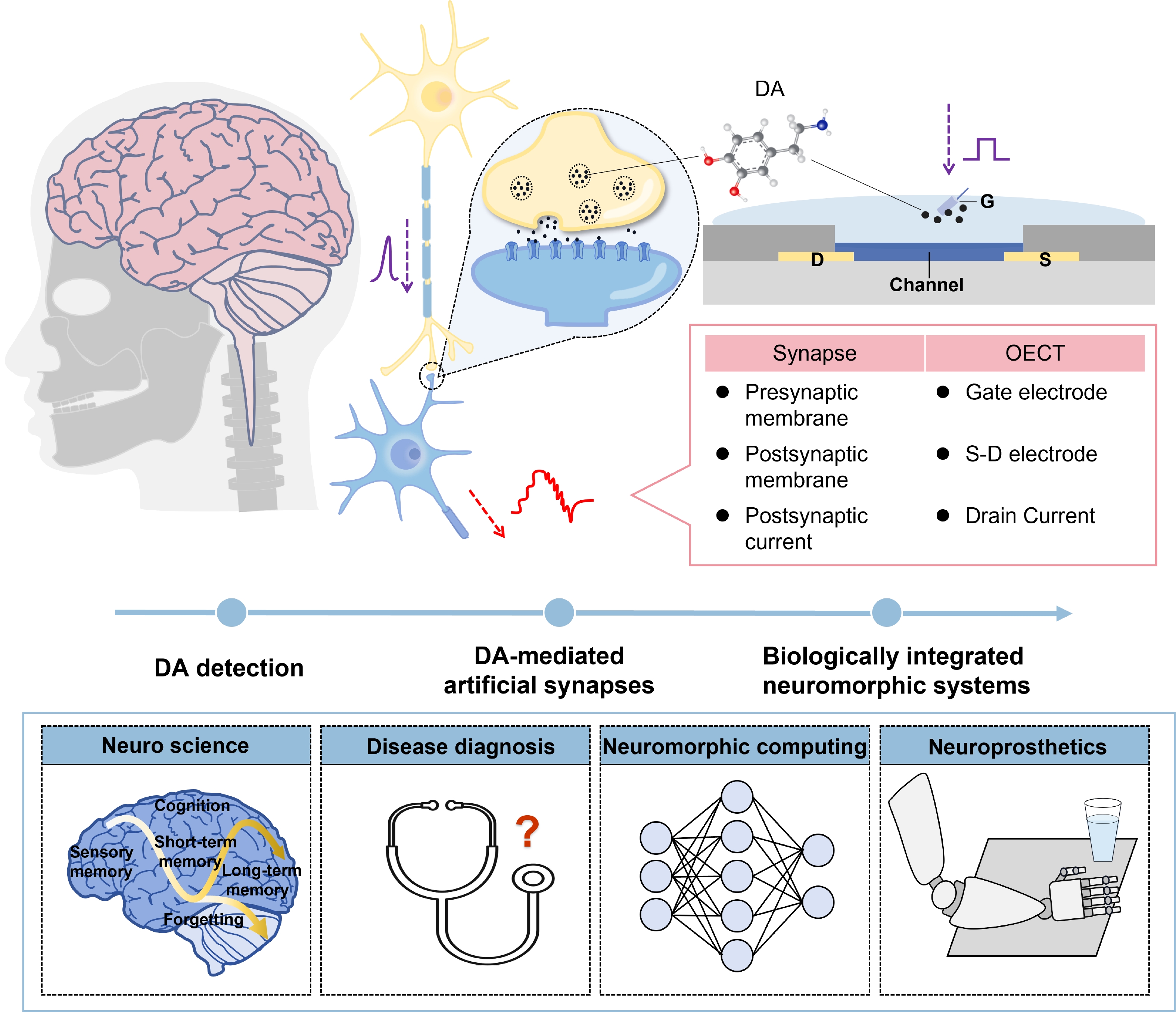
Organic electrochemical transistors (OECTs) are electronic devices that rely on organic mixed ionic–electronic conductors (OMIECs) and can operate in aqueous environments[15]. Additional advantages of OECTs include low operating voltage, good biocompatibility, and high transconductance[16, 17], making them ideal platforms for interfacing with biological systems for biochemical and electrophysiology sensing. These attributes are particularly beneficial in biochemical sensors, point-of-care diagnostics, and implantable and wearable technologies[1824]. Note that OECTs possess a similar structure to biological synapses as shown in Fig. 1, with channels forming the synaptic gap of artificial chemical synapses. The gate electrode corresponds to the presynaptic membrane, which receives the stimulus, and the drain electrode resembles the postsynaptic membrane, collecting the postsynaptic current (PSC) for further signal processing[25]. Therefore, OECTs are an ideal platform for biomimic neurotransmitter-synapse interactions.

Fig. 1.  (Color online) Concept of artificial synapses with neurotransmitters-mediated functions. OECTs structures are similar to biological synapses, and neurotransmitter-mediated artificial synapses have potential implications in the fields of neuroscience, disease diagnosis, neuromorphic computing, and neuroprosthetics.

While several good review articles related to artificial synapses[18, 21, 2629] and bioelectronic interfaces[19, 23, 30, 31] have been published recently, there is a notable lack of comprehensive summaries specifically on artificial synapses mediated by neurotransmitters and resultant neuromorphic systems, which are key for biomimic and bioinspired neuromorphic systems. This review article aims to fill this gap. In the first part of this contribution, the device structure and operational mechanism of OECTs are introduced. The second part focuses on the advances on the detection of a representative neurotransmitter, dopamine (DA), which can modulate synaptic transmission between neuros and thereby influence synaptic conditioning and plasticity. The detection principles and methods to improve sensor performance as well as in vivo detection are also summarized. The third part discusses the neurotransmitter-mediated artificial synapses, including synaptic plasticity, and examples related to simulation of biological synapses and biologically integrated neuromorphic systems. Finally, the possible prospects and future research directions for future biologically integrated neuromorphic systems through neurotransmitter-mediated artificial synapses are proposed.

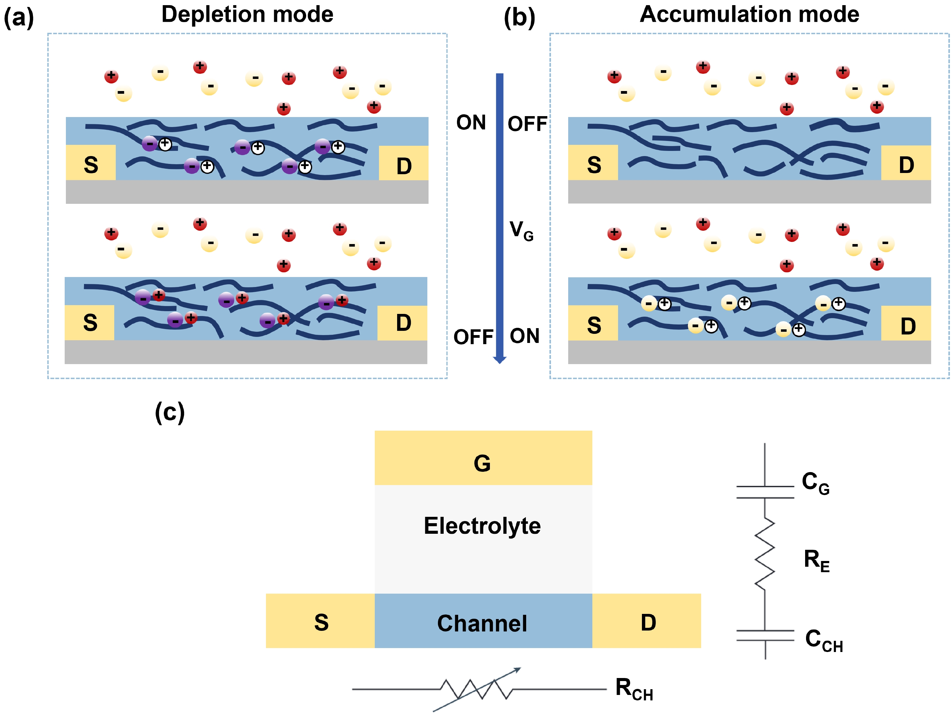
OECTs are three-terminal devices, with a drain, source and gate electrodes. Same with metal−oxide semiconductor field-effect transistors (MOSFETs) and organic field-effect transistors (OFETs), OECTs can be considered as amplifiers or switches, where the input signal (VG) amplifies and controls the output signal (ID). Distinct from OFETs, the gate electrode of OECTs is isolated from the channel through the use of an electrolyte. The electrolyte can be a liquid, solid, or gel[15, 32, 33]. The channel layer in OECTs is an OMIEC, which is both electronically and ionically conductive, and the electrolyte with ionic conductivity can promote the movement of ions under the action of an electric field. OECTs can be divided into two types according to different working states: 1) The channel material conducts electricity and typically operates in depletion mode (ON to OFF), and the input (VG) causes electrochemical dedoping, thereby reducing the electrical conductivity of the channel (Fig. 2(a)). 2) The channel material is poorly conductive and operates in the accumulation mode (OFF to ON), and its doping requires the application of a gate voltage (VG), thereby increasing the conductivity of the channel (Fig. 2(b)). The most popular semiconductor material in OECTs is poly(3,4-ethylenedioxythiophene):poly(styrene sulfonate) (PEDOT:PSS), which operates in the depletion mode[22].

Fig. 2.  (Color online) Mechanism of OECTs. (a) Depletion and (b) accumulation mode of OECTs. (c) Two circuits (electronic and ionic circuits) used to model OECTs.

According to Bernards' model[34], OECT can be divided into two circuits (Fig. 2(c)). The first is the electronic circuit, modeled as a resistor (RCH), which considers the movement of charge carriers within the source−channel−drain structure. The second is the ionic circuit in the gate-electrolyte-channel configuration. A resistor (RE), gate capacitor (CG) and channel capacitor (CCH) were involved. As a main figure of merit, the transconductance (gm) of OECTs is defined as the ratio of the change in ID to the change in VG, and can be expressed as (Eq. (1)):

gm=IDVG=WdLμC(VTVG),
(1)

where d is the channel thickness, L is the channel length, W is the channel width, C* is the volumetric capacitance, VT is the threshold voltage, and μ is the charge carrier mobility. Increasing W and decreasing L can effectively enhance gm, but increasing d is a more practical approach for the gm improvement due to the challenge of device miniaturization[35, 36]. Note that a thicker channel layer typically leads to a slower response time for OECTs because of the longer pathways for ion penetration. Another method for gm improvement is to design and synthesize new channel materials with higher μC*[37, 38]. Besides, switching times, on/off current ratio (Ion/Ioff), and stability also need to be considered to comprehensively evaluate the performance of OECTs[39, 40].

The working principle of the DA sensor based on OECTs is shown in Fig. 3(a). Within the electrolyte, DA molecules can undergo electro-oxidation in the form of o-dopaminequinone at the gate electrode, and therefore a faradic current is generated[41]. The advantages of high transconductance, low operating voltage and good aqueous compatibility allow OECTs amplifying the weak signal of DA in solution. This process decreases the potential drop at the electrolyte/gate interface and increases the effective gate voltage (Vg-eff) (Fig. 3(b)), as detailed by[42]:

Fig. 3.  (Color online) Working principle of OECTs for DA detection. (a) Schematic of DA oxidation on the OECT gate surface. The electrodes can be modified by nanoparticles (NPs), carbon materials, aptamers, molecularly imprinted polymers (MIPs), etc. (b) Potential changes in OECTs. (c) ID changed over time as DA was added[41]. Reproduced with permission. Copyright 2011, Elsevier. (d) Curve of ΔVg-eff versus change in DA concentration[41]. Reproduced with permission. Copyright 2011, Elsevier.
ΔVg-eff={2.30(1+γ)kT2qlog[C]higher concentrationA×[C]βlow concentration,
(2)

where [C] is the DA concentration, T is the absolute temperature, k is the Boltzmann constant, q is the charge of an electron, A and β are constants determined by fitting experimental data, γ is the ratio between the volumetric capacitance of the channel (CCH) and the capacitance of the gate-electrolyte interface (CG). In the higher concentration range, the potential ΔVg-eff will increase linearly with the increase in the logarithm of [C]. However, in the low concentration range (below nM), the nonlinear relation was often observed[41]. Therefore, Zhang et al. proposed an empirical relationship in which ΔVg-eff is power-exponential to [C][43]. In practice, the sensitivity of the device at low concentrations needs to be calibrated.

The real-time ID changes of an OECT device during adding different concentrations of DA into phosphate buffer saline is shown in Fig. 3(c), with the fitted calibration curve (Fig. 3(d)). The sensor performance can be evaluated by two key parameters that are extracted from the calibration curve: the sensitivity and the limit of detection (LOD). The sensitivity defines the minimum input required to generate a detectable output response from the sensor. The most common unit of sensitivity is mV/dec, while some reports also used current change to describe the sensitivity with a unit of mA/dec or the gm change with a unit of SM−1[44, 45], while the LOD indicates the lowest analyte concentration that can be reliably detected compared to a blank measurement. In this detection principle, selectivity is defined as the ratio of the slope of the calibration line for the analyte of interest to the slope of the specific interferent. However, in practical measurements, less attention is paid to this parameter[46]. Another parameter that needs to be considered is the linear range of DA detection, where the output is proportional to the input. Within this range, the sensitivity is theoretically maintained at a constant level, thereby enabling the sensor to ensure a certain level of measurement accuracy as the range increases. The greater the linear range of the sensor, the larger its detection range, which is essential for ensuring measurement accuracy[47].

From Eq. (3), ΔVg-eff is proportional to log[C]. The slope of the fitted curve can be modulated by controlling the capacitance ratio γ[41]:

γ=CCHCG=cchAchcgAg,
(3)

where AG and ACH are the areas of gate and channel electrode and cg and cch are the gate and channel capacitances per unit area, respectively. The capacitance ratio is proportional to ACH/AG. Therefore, optimizing the ratio of the channel area to the gate area in OECTs can enhance the performance of DA detection. Additionally, it has been reported that alternating current (AC) methods offer greater sensitivity and more stable data in sensing applications compared to traditional direct current (DC) methods[48].

DA is an electrically active molecule that can be oxidized without the enzymes, and therefore it can be quantified through the electrochemistry technique. In particular, the oxidation peak of DA is 150 mV (compared to Ag/AgCl)[46]. However, a few challenges still exist. On one hand, most reported electrochemical DA sensors exhibited linear detection ranges from 1 to 100 mM with the detection limits around 1 mM[4951], which are orders of magnitude higher than the DA concentrations in clinic samples (nM for plasma, μM for urine and fM for single adrenal chromaffin cell, respectively[52, 53]). On the other hand, DA has many analogues, and a number of potential interferences like ascorbic acid (AA) and uric acid (UA) also complicate the specific detection[46, 54]. Interestingly, the high transconductance of OECTs allows sufficient signal amplification and consequent detection limit of DA down to the nM, suggesting the detection capability in real samples like urine and plasma[41]. OECTs-based DA sensors have a faster response time compared to conventional methods that typically require several minutes of response time, often within seconds of detection[55, 56]. Besides, the specificity for DA sensing can be improved by modifying various active materials on the gate of OECTs. Table 1 summarizes the characteristics and structures of recent DA detectors based on OECTs.

Table 1.  Comparisons of different OECT-based DA biosensors.
Gate materialsGate area (μm2)Channel size
(W/L (μm/μm))
LOD (M)Linear range (M)Sensitivity
(mV/dec)
Ref.
Aptamer-enhanced Au/30/225 × 10−165 × 10−12−10−9/[57]
9 × 10⁶/1 × 10−1410−11−10−86.9 [58]
Pt9 × 10622/51 × 10−9 (AC)
1 × 10⁻⁸ (DC)
10−7−10−545.2[48]
4.8 × 10540/103 × 10−83 × 10−8−1 × 10⁻⁷/[42]
/6000/100<5 × 10−95 × 10−8 −3 × 10−6174[41]
Vertical Pt wire2.8 × 105700/19004.4 × 10−6/410[59]
Nafion and graphene flakes co-modified Pt/6000/2005 × 10−95 × 10−9−1 × 10−6242[60]
Overoxidized MIP (o-MIP/Pt)/1000/203.4 × 10−84 × 10−7−1 × 10⁻⁵/[61]
Ag/AgCl pseudo reference/10/10/1 × 10−6−3 × 10−4279[62]
/5000/15504.3 × 10−81 × 10−9−1 × 10−40.312 mA/dec[44]
/1500/50003.7 × 10−85 × 10−8−1 × 10⁻⁴/[63]
Nitrogen/oxygen-codoped carbon cloths//1 × 10−91 × 10−6−3 × 10−4151[39]
Carbon/PEDOT: PSS3.9 × 10−20.45 × 0.45 × π (needle-typed)1 × 10−121 × 10−9−1.6 × 10⁻⁷
2 × 10⁻⁹−7 × 10⁻⁶
190 ± 20 pA/dec
210 ± 40 pA/dec
[64]
CNT/ (Pt NPs) fiber//5 × 10−95 × 10−9−5 × 10⁻⁷/[65]
rGO/CNT/PEDOT:PSS/240/776 × 10−61 × 10−5−1 × 10⁻⁴/[66]
Polypyrrole nanofiber//1 × 10−91 × 10−9− 1 × 10⁻⁶/[67]
PEDOT:PSS9 × 1063000/30006 × 10−65 × 10−6−1 × 10⁻⁴/[68]
Copper phthalocyanine/166/203.04 × 10−65 × 10−5−5 × 10⁻⁴/[16]
DownLoad: CSV  | Show Table

By employing various gate electrodes (gold, graphite, and platinum (Pt)), Tang et al.[41] realized DA detection using OECTs. It was found that the OECT sensitivity were dependent on the gate electrode's characteristics and the operational voltage. Due to the excellent catalytic properties of Pt, the device with a Pt electrode at a gate voltage of 0.6 V had the highest sensitivity. Moreover, a LOD of less than 5 nM for DA was achieved, indicating a significant improvement over conventional electrochemical methods with the same electrode. But this sensor had poor selectivity and sensitivity. Several research groups have also used Ag/AgCl pseudo reference electrode[44, 62, 63] as gate electrode. Although they could achieve good performance due to unpolarizable property[38], metal electrodes were more advantageous in terms of integration. Thereafter, a wide variety of DA sensors based on OECTs were developed through gate modification and architecture design.

Modifying the gate electrode with various materials in OECTs can effectively enhance both the selectivity and sensitivity for DA detection. These modifications help reduce interference and get a lower LOD. Liao et al.[60] coated the Pt gate surface with a biocompatible polymer Nafion or chitosan, by which the interference induced by UA and AA was effectively eliminated, particularly after the modification with Nafion. Additionally, the incorporation of graphene flakes on the gate electrodes further increased the sensitivity, achieving a DA detection limit of 5 nM. This value was significantly lower than that of conventional electrochemical methods. Besides, widely explored nanomaterials such as gold NPs[63], platinum NPs[65], or various composite materials[67] were also sufficient to enhance the performance of OECTs-based DA sensors as the modification layer.

Different from commonly used planar device structure, Mariani et al.[64] designed a needle-type OECT for spatially resolved detection of DA. Due to its unique structure, the sensor featured a high gate-to-channel area ratio. Besides, this study combined the advantages of carbon nanoelectrodes (CNEs) with OECTs to realize nanoscale pioneer OECTs, where both single- and double-tube structures were used with a conductive polymer film on the CNEs. Intriguingly, this needle-type OECT had a size similar with a single cell, and therefore high spatial resolution detection of neurotransmitters can be realized. By applying appropriate drain and gate voltages, this OECT sensor achieved a LOD on the order of pM and a linear DA detection range from 2 nM to 7 μM. Other OECT architecture such as self-curling[16] and fiber-shaped structures[59, 65, 67] were also proposed with good detection performance.

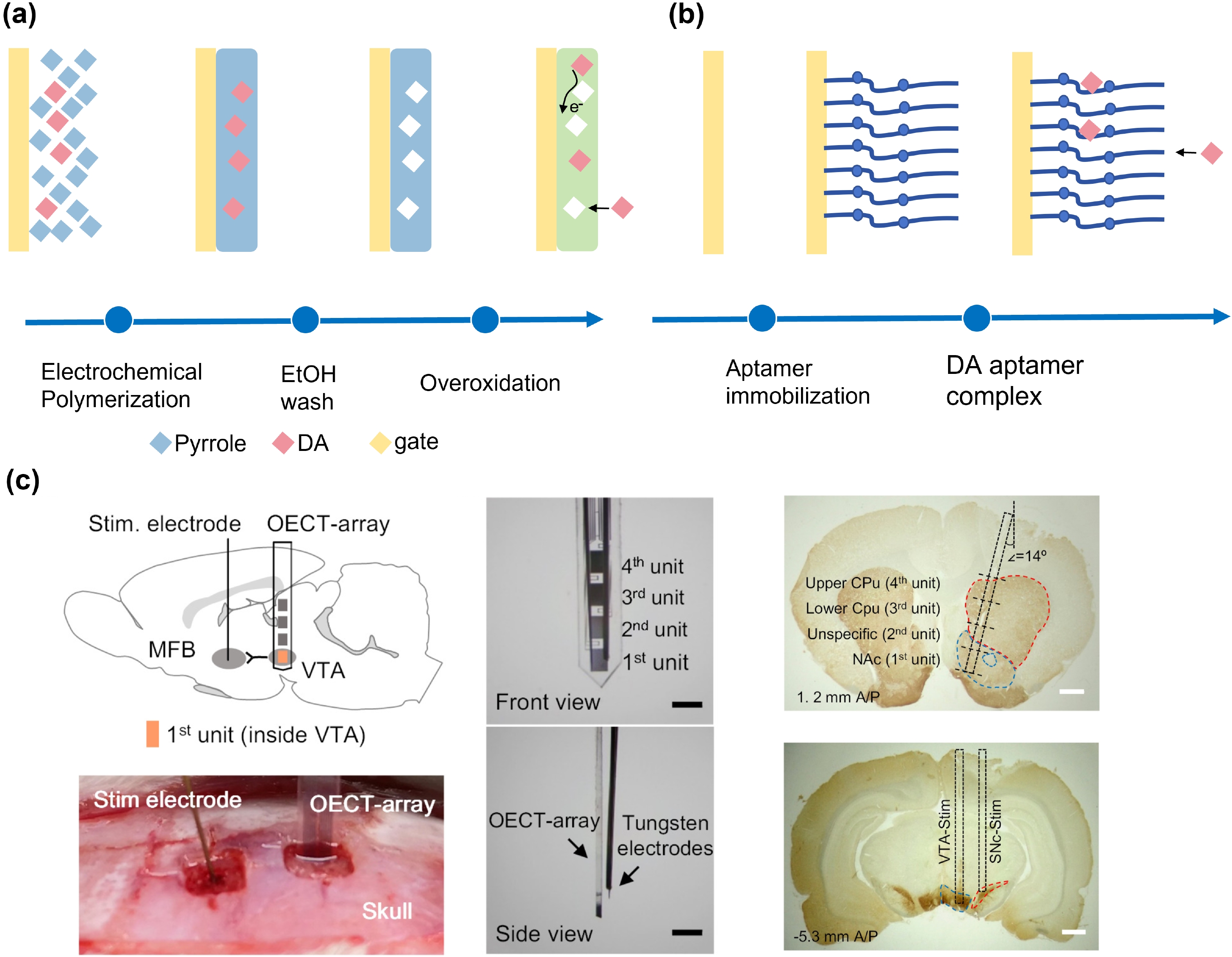
MIPs[69] are useful materials for selective binding, where a template molecule (target) is dissolved in a suitable solvent with one or more monomers and a cross-linking agent to form a highly cross-linked polymer. Following polymerization, the template molecules are removed, and consequently nanocavities are formed that correspond to the size, shape, chemical interactions and orientation of the template molecules[70]. Therefore, the gate modification using MIPs was found to be an effective approach for high-performance OECT DA sensor[61]. The flow of electrochemical synthesis of overoxidized MIP (o-MIP) on OECT gate is shown in Fig. 4(a).

Fig. 4.  (Color online) (a) Electrochemical synthesis of DA-specific o-MIP at gate. (b) Operation principle of aptamer immobilization. (c) Real-time detection of neurotransmitter released in rat brain by OECT array. Reproduced with permission[42]. Copyright 2020, eLife Sciences Publications, Ltd.

The OECT sensor with an o-MIP/Pt gate showed a LOD of 34 nM, which is almost identical to the one with bare Pt. However, the selectivity was remarkably improved, and a DA/AA signal ratio over 5 ranging from ~0.4 to ~10 μM was obtained.

Aptamers are a superior option for DA sensors, irrespective of cost considerations. Aptamers, which are made up of single-stranded RNA or DNA molecules, have the ability to bind detection molecules with outstanding affinity and specificity[71]. In contrast to large antibodies, aptamers typically have a lower molecular weight. In addition, aptamers can be used to develop biosensors with specificity and fast response due to their conformational changes upon binding. A considerable number of naturally occurring aptamers have been identified in the untranslated regions of mRNA. These aptamers have been demonstrated to bind small molecule metabolites[71, 72]. As a result, many efforts have been made to extract DA aptamers and have been used in sensors[46, 57, 73, 74].

Soliman et al.[58] developed aptamer-enhanced OECTs for ultra-sensitive DA detection. Fig. 4(b) illustrates the operational principle of aptamer immobilization in conjunction with an integrated OECT with a functionalized gate. The sensor demonstrated real-time detection with a linear response from 10 fM to 10 nM and a detection limit of 10 fM. In addition, Liang et al. reported similar aptamer sensor, where a fM level DA detection was also obtained with high sensitivity[57]. This provides an efficient platform for future disease diagnosis and clinical applications. However, more efforts are needed to ensure reusability and versatile integration with other devices.

Real-time in vivo detection of DA is necessary to build integrated bio-logical neuromorphic systems. However, the complex environments that usually accompany in-vivo situations in living organisms require the fabrication of more well-designed structure with higher sensitivity and stability[11]. This can be addressed by using OECT devices. Li et al.[75] demonstrated a fast-scanning potential (FSP)-gated OECT, where a sensitivity of 0.899 SM−1 and a LOD of 5 nM were reported. Importantly, the real-time dopamine monitoring was realized using this device in the living rat brain. Unlike the previous individual devices, Xie et al.[42] fabricated an OECT array for monitoring DA in rat brains (Fig. 5(c)). The OECT array consists of multiple OECTs that can detect transient DA releases in real time with nanomolar scale LOD and high temporal resolution. Notably, continuous in vivo operation for several hours was reported at an operating voltage under 1 V without significant degradation. Additionally, these OECT arrays were capable of simultaneously detecting evoked DA release in multiple striatal brain regions.

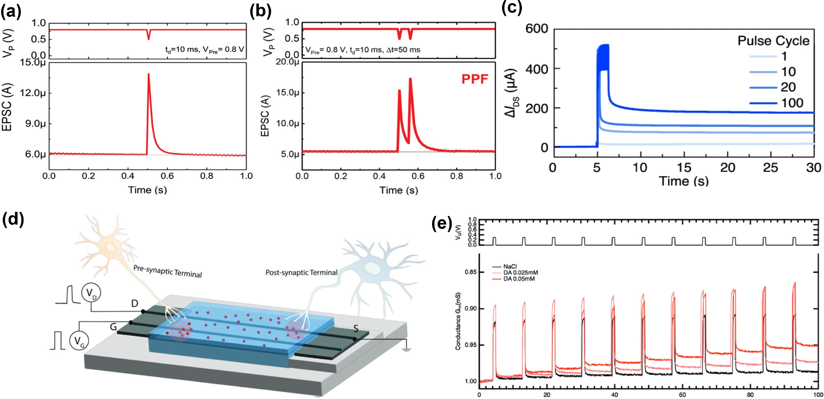
Fig. 5.  (Color online) Synaptic plasticity of OECTs. (a) Excitatory postsynaptic current (EPSC)[76]. Reproduced with permission. Copyright 2019, Wiley. (b) Paired-pulse facilitation (PPF, A2/A1)[76]. Reproduced with permission. Copyright 2019, Wiley. (c) Retention curves for synaptic weight after different number of applied pulses, showing STP−LTP[77]. Reproduced with permission. Copyright 2021, Springer Nature. (d) and (e) An artificial fiber bio-hybrid synapse with DA-mediated plasticity[78]. Reproduced with permission. Copyright 2024, Wiley.

Synaptic plasticity, a biological process, is defined as the alteration of synaptic weight under certain conditions. It is regarded as a fundamental mechanism that underlies the ability of the brain to adapt and learn[79]. Simulating synaptic plasticity through OECTs is a prerequisite for constructing ANNs and SNNs. In addition, neurotransmitters are important for synaptic plasticity. In response to the activation of receptors by a variety of neurotransmitters, chemical synapses amplify and transform presynaptic signals, which in turn have a complex effect: excitatory postsynaptic potential (EPSP) or inhibitory postsynaptic potential (IPSP) on the postsynaptic neuron[28].

Based on the duration of synaptic weight changes, synaptic plasticity can be categorized into two distinct types: short-term (STP) and long-term plasticity (LTP). Taking the neuronal excitation process as an example, a short single presynaptic voltage spike triggers a sharp increase in excitatory postsynaptic current (EPSC) to a level, A1, which then quickly returns to its baseline. This behavior is called STP (Fig. 5(a))[76]. If a subsequent voltage pulse is applied before the EPSC has fully decayed, the current A2 is higher than A1. This behavior is defined as paired-pulse facilitation (PPF), and its strength is quantified as A2/A1 (Fig. 5(b))[76]. LTP occurs in response to a high frequency of presynaptic action potentials. With increasing the number of presynaptic spikes applied to the organic synapse, a transition of synaptic response from STP to LTP is possible (Fig. 5(c))[77, 80]. During LTP, the synaptic weight increases, and then decays slowly to a certain state that maintains for a long time.

Achieving synaptic modulation based on biochemical signaling detection is the first step towards a biologically integrated neuromorphic system[12]. Significant contributions to devices that simulate DA recognition and release have been made, bridging the connection between biological neural networks and artificial intelligence systems[81, 82]. The neuromorphic response induced by the redox reaction of DA is one of the most efficient ways to realize this process. Table 2 summarizes the DA-mediated artificial synapse based OECTs.

Table 2.  Comparisons of DA-modulated artificial synapses based-OECTs.
TypeGate materialsKey propertiesDetailsRef.
Biohybrid synapseAu/PEDOT: PSSDA recycling/LTP/LTDOECT/microfluidic channel[12]
PEDOT: PSS fiberDA recyclingFiber OECT[78]
Artificial spiking neuronAg/AgClDA/ions sensitive and neuron spiking propertiesOECT[85]
AuDA/5-HT sensitive and neuron spiking properties/ biological functions of a retinal pathwayOECT/microfluidic channel[86]
Artificial synapsesAg ink
STP/LTP/flexibleFully printed dual-gate OECT[83]
ITOMimicking biological neurotransmitter coreleaseElectrical and optical modulation OECT/microfluidic channel[84]
Closed-loop neuromorphic systemPEDOT: PSSClosed-loop control, actuation and reinforcement learningOECT/microfluidic channel/robotic hand[14]
DownLoad: CSV  | Show Table

Keene et al.[12] demonstrated a functional biohybrid synapse, in which a dopaminergic presynaptic domain of PC-12 cells was coupled to an organic neuromorphic device serving as the postsynaptic domain. This design allowed OECTs as artificial synapses direct contact with living tissue and adaption through biofeedback. Furthermore, the electrochemical simulation of long-term potentiation and long-term depression of synaptic weights induced by DA were also achieved. Additionally, polydimethylsiloxane (PDMS) microfluidic channels allowed for precise control of fluid flow within the device, which mimicked DA endocytic recycling of in biological synapses. Subsequently, several successive studies[78, 83, 84] have investigated artificial synapses with DA-mediated plasticity based on OECTs. DA-mediated plasticity is markedly distinct from the short-term synaptic behavior observed in electrolytes devoid of DA. The rationale for this phenomenon can be elucidated by the chemical reaction where DA is oxidized to DA o-quinone in the presynaptic compartment, resulting in the simultaneous production of 2e and 2H+. These generated H+ ions then inserted into the channel, where they combined with PSS to form stable sulfonic acid groups, thereby facilitating LTP[83].

The device structure is important in neuromorphic devices. Fibers are lightweight, flexible, breathable and comfortable compared to thin-film structures[87, 88]. These characteristics confer a significant advantage in the field of wearable technology and BCIs. Alarcon-Espejo et al.[78] developed the fiber-electrochemical neuromorphic organic devices (Fiber-ENODes), and demonstrated that these high hole mobility fibers hold great potential as foundational elements for future bio-hybrid technologies. Fig. 5(d) showed the structure of artificial bio-hybrid synapse based on double PEDOT: PSS fiber. It was revealed that the optimization of electrode/organic interface and fiber alignment resulted in stable contacts, high μC* products, and high hole mobility. The device respond to DA was found to affect STP and LTP for Fiber-ENODes. Moreover, a shape factor very similar to neuronal axon terminals was also observed, as shown in Fig. 5(e).

BCI technology is a transformative human-computer interaction technology[31, 89]. Its mechanism of action is to bypass peripheral nerves and muscles and directly establish a brand new communication and control channel between the brain and external devices. Artificial synapses based on OECTs are expected to accelerate the development of BCI technology as a bridge between machines and living organisms, thereby enabling biologically integrated neuromorphic systems. Currently the detection of electrical signals in organisms has been successfully realized by OECTs[20, 90, 91]. In addition to electrical signals, chemical signals are also a crucial part of living organisms[92]. Therefore, artificial synapses with neurotransmitter-mediated is of great importance in future applications of BCIs.

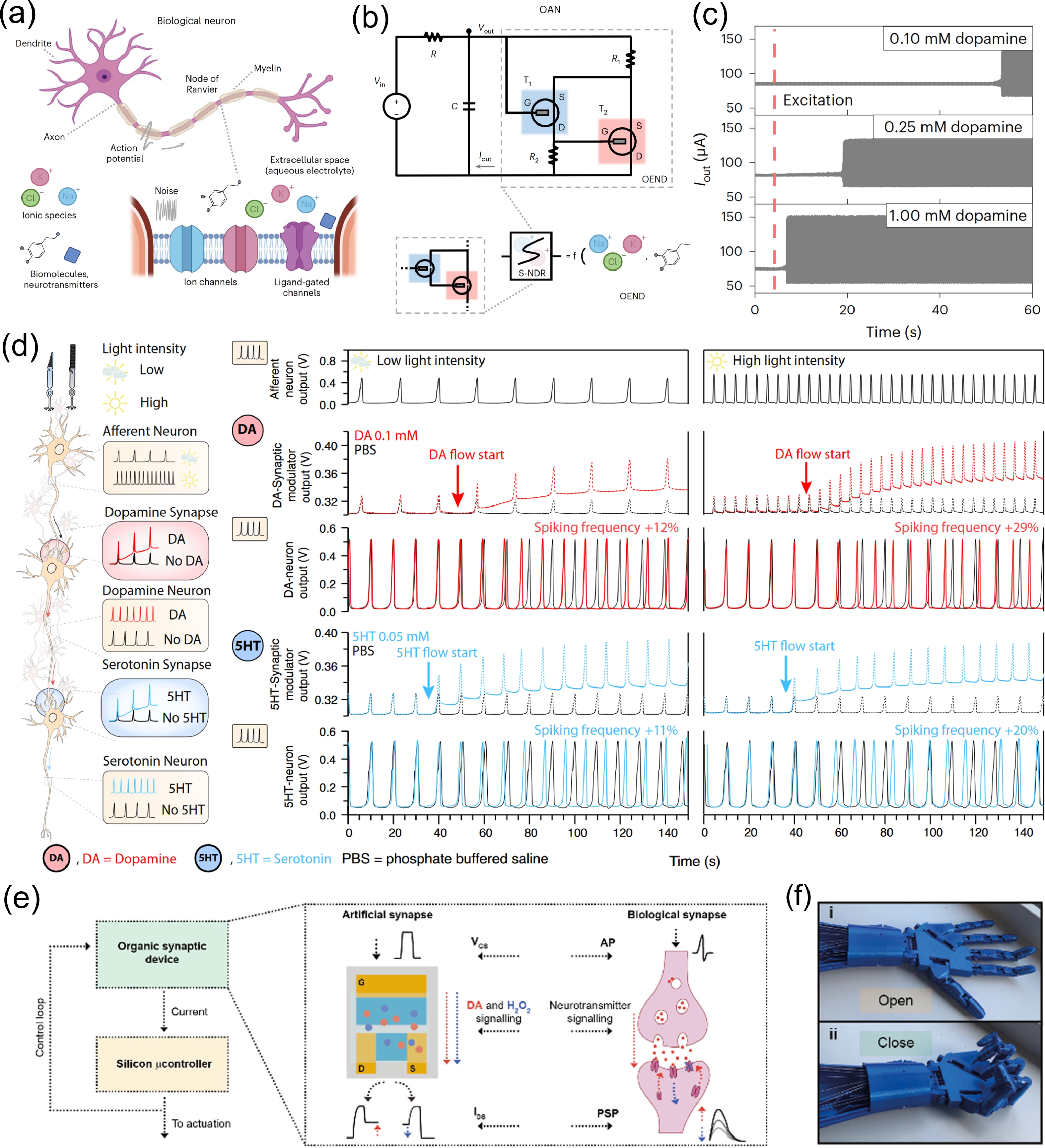
Sarkar et al.[85] fabricated an organic artificial neuron (OAN) using a compact nonlinear electrochemical element. This device with inherent biosensing capabilities could operate in liquid environments, as shown in Figs. 6(a) and 6(b), and more importantly, mimics the spiking behavior of neurons. Its sensitivity to ion concentration and dopamine concentration was confirmed, facilitating the modulation of spiking excitatory behavior (Fig. 6(c)). Matrone et al. realized a more complex system[86]. This system used OANs to mimic afferent neurons that received light stimulation and employed synapses modulated by DA and Serotonin (5-HT) to simulate the neuromodulatory activity of intermediate neurons. As shown in the Fig. 6(d), an OECT synapse can modulate the spike pulses from the preceding neuron based on DA concentration. As concentration increased, the synaptic output significantly enhanced, leading to an increase in the spike pulse frequency of the subsequent neuron. The degree of modulation depended on the concentration of the neurotransmitter. This process effectively simulated the spike-rate coding dependent on neurotransmitters in intermediate neurons and the transmission of neural signals between multiple neurons.

Fig. 6.  (Color online) Biologically integrated neuromorphic systems. (a) and (b) A biological neuron and the OAN. Reproduced with permission[85]. Copyright 2022, Springer Nature. (c) The OAN is sensitive to DA and exhibits neuronal spiking properties and excitability modulation. Reproduced with permission. Copyright 2022, Springer Nature. (d) Neuromorphic artificial retinal pathways combining sensory coding and neuromodulation[86]. Reproduced with permission. Copyright 2024, Springer Nature. (e) and (f) Closed-loop neuromorphic system and robotic actuation[14]. Reproduced with permission. Copyright 2024, Royal Society of Chemistry.

In order to control the machine by the organisms, Bruno et al. designed an OECT platform through neurotransmitter closed-loop control, actuation and reinforcement learning[14]. The overall neuromorphic closed-loop architecture was shown in Fig. 6(e), where the standard silicon technologies were utilized. This design enabled brain-inspired computing through adaptive synaptic potentiation and depression within a closed-loop system. Specifically, it utilized DA release and detection to facilitate closed-loop learning and training processes akin to those in the human brain. Notably, a robotic hand could be interfaced and controlled by this microfabricated platform, as shown in Fig. 6(f), and autonomous learning to grasp objects of different sizes was also realized. Many efforts have been made by researchers to realize bio-integrated neuronal networks. However, the field is still in its infancy.

In recent years, the development of OECT technology and the trend of multidisciplinary intersection have facilitated the rapid development of OECT-based DA sensors. In this review, an overview of the structure and operating mechanism of OECTs is first presented. Then a comprehensive review of the advances in DA sensors and artificial synapses based on OECT technology is discussed. By combining DA detection and modulation, artificial synapses enable chemical communication with neurotransmitter interactions. The platform has significant implications for neuroscience, disease diagnosis, neuroprosthetic control, bioelectronic medicine, and the integration of brain and machine intelligence. However, many challenges remain to be addressed. The first problem faced is how to reduce power consumption and improve long-term stability. As more devices are integrated, reducing power consumption to levels comparable to that of the biological brain is becoming increasingly critical. Furthermore, the working life on a yearly basis is required for the implantable devices, and therefore the long-term stability of current OECTs should be significantly improved. The second is to increase the diversity of sensing. While considerable progress has been made in DA detection, synaptic events are usually the result of multiple neurotransmitters acting together[93, 94]. Therefore, there is a high demand to develop sensors that can detect multiple neurotransmitters simultaneously. The third is to achieve a closed loop of OECTs and neurotransmitter signaling. OECTs can simulate synaptic plasticity, spike modulation and adaptive learning, but integration with biological systems requires the bidirectional communication[95]. This requires the use of BCI technology to create a closed loop between the artificial synapse and the organism.

This work is supported by the National Natural Science Foundation of China (Grant No. 62074163) and Beijing Natural Science Foundation (Grant No. JQ24030).


[1]
Pereda A E. Electrical synapses and their functional interactions with chemical synapses. Nat Rev Neurosci, 2014, 15, 250 doi: 10.1038/nrn3708
[2]
Smart T G, Paoletti P. Synaptic neurotransmitter-gated receptors. Cold Spring Harb Perspect Biol, 2012, 4, a009662 doi: 10.1101/cshperspect.a009662
[3]
Wang T, Wang M, Wang J W, et al. A chemically mediated artificial neuron. Nat Electron, 2022, 5, 586 doi: 10.1038/s41928-022-00803-0
[4]
Hyman S E. Neurotransmitters. Curr Biol, 2005, 15, R154 doi: 10.1016/j.cub.2005.02.037
[5]
Zeisel S H. A brief history of choline. Ann Nutr Metab, 2012, 61, 254 doi: 10.1159/000343120
[6]
Kelly R B. Storage and release of neurotransmitters. Cell, 1993, 72, 43 doi: 10.1016/S0092-8674(05)80027-3
[7]
Su Y, Bian S M, Sawan M. Real-time in vivo detection techniques for neurotransmitters: A review. Analyst, 2020, 145, 6193 doi: 10.1039/D0AN01175D
[8]
Frieling H, Hillemacher T, Demling J, et al. Neue wege in der depressionsbehandlung. Fortschr Neurol Psychiatr, 2007, 75, 641 doi: 10.1055/s-2007-959181
[9]
Pan J X, Xia J J, Deng F L, et al. Diagnosis of major depressive disorder based on changes in multiple plasma neurotransmitters: A targeted metabolomics study. Transl Psychiatry, 2018, 8, 130 doi: 10.1038/s41398-018-0183-x
[10]
Iversen S D, Iversen L L. Dopamine: 50 years in perspective. Trends Neurosci, 2007, 30, 188 doi: 10.1016/j.tins.2007.03.002
[11]
Banerjee S, McCracken S, Hossain M F, et al. Electrochemical detection of neurotransmitters. Biosensors, 2020, 10, 101 doi: 10.3390/bios10080101
[12]
Keene S T, Lubrano C, Kazemzadeh S, et al. A biohybrid synapse with neurotransmitter-mediated plasticity. Nat Mater, 2020, 19, 969 doi: 10.1038/s41563-020-0703-y
[13]
Krauhausen I, Griggs S, McCulloch I, et al. Bio-inspired multimodal learning with organic neuromorphic electronics for behavioral conditioning in robotics. Nat Commun, 2024, 15, 4765 doi: 10.1038/s41467-024-48881-2
[14]
Bruno U, Rana D, Ausilio C, et al. An organic brain-inspired platform with neurotransmitter closed-loop control, actuation and reinforcement learning. Mater Horiz, 2024, 11, 2865 doi: 10.1039/D3MH02202A
[15]
Rivnay J, Inal S, Salleo A, et al. Organic electrochemical transistors. Nat Rev Mater, 2018, 3, 17086 doi: 10.1038/natrevmats.2017.86
[16]
Ferro L M M, Merces L, de Camargo D H S, et al. Organic electrochemical transistors: Ultrahigh-gain organic electrochemical transistor chemosensors based on self-curled nanomembranes. Adv Mater, 2021, 33, 2170223 doi: 10.1002/adma.202170223
[17]
Khodagholy D, Rivnay J, Sessolo M, et al. High transconductance organic electrochemical transistors. Nat Commun, 2013, 4, 2133 doi: 10.1038/ncomms3133
[18]
Liu H, Song J J, Zhao Z Y, et al. Organic electrochemical transistors for biomarker detections. Adv Sci, 2024, 11, 2305347 doi: 10.1002/advs.202305347
[19]
Spanu A, Martines L, Bonfiglio A. Interfacing cells with organic transistors: A review of in vitro and in vivo applications. Lab Chip, 2021, 21, 795 doi: 10.1039/D0LC01007C
[20]
Uguz I, Ohayon D, Arslan V, et al. Flexible switch matrix addressable electrode arrays with organic electrochemical transistor and pn diode technology. Nat Commun, 2024, 15, 533 doi: 10.1038/s41467-023-44024-1
[21]
Nawaz A, Liu Q, Leong W L, et al. Organic electrochemical transistors for in vivo bioelectronics. Adv Mater, 2021, 33, 2170387 doi: 10.1002/adma.202170387
[22]
Marks A, Griggs S, Gasparini N, et al. Organic electrochemical transistors: An emerging technology for biosensing. Adv Mater Interfaces, 2022, 9, 2102039 doi: 10.1002/admi.202102039
[23]
Saleh A, Koklu A, Uguz I, et al. Bioelectronic interfaces of organic electrochemical transistors. Nat Rev Bioeng, 2024, 2, 559 doi: 10.1038/s44222-024-00180-7
[24]
Li N, Li Y, Cheng Z, et al. Bioadhesive polymer semiconductors and transistors for intimate biointerfaces. Science, 2023, 381, 686 doi: 10.1126/science.adg8758
[25]
Chouhdry H H, Lee D H, Bag A, et al. A flexible artificial chemosensory neuronal synapse based on chemoreceptive ionogel-gated electrochemical transistor. Nat Commun, 2023, 14, 821 doi: 10.1038/s41467-023-36480-6
[26]
Dai S L, Zhao Y W, Wang Y, et al. Recent advances in transistor-based artificial synapses. Adv Funct Mater, 2019, 29, 1903700 doi: 10.1002/adfm.201903700
[27]
Huh W, Lee D H, Lee C H. Memristors based on 2D materials as an artificial synapse for neuromorphic electronics. Adv Mater, 2020, 32, 2002092 doi: 10.1002/adma.202002092
[28]
Bhunia R, Boahen E K, Kim D J, et al. Neural-inspired artificial synapses based on low-voltage operated organic electrochemical transistors. J Mater Chem C, 2023, 11, 7485 doi: 10.1039/D3TC00752A
[29]
Han H, Yu H Y, Wei H H, et al. Recent progress in three-terminal artificial synapses: From device to system. Small, 2019, 15, 1900695 doi: 10.1002/smll.201900695
[30]
Gkoupidenis P, Zhang Y, Kleemann H, et al. Organic mixed conductors for bioinspired electronics. Nat Rev Mater, 2024, 9, 134 doi: 10.1038/s41578-023-00622-5
[31]
Tang X, Shen H, Zhao S Y, et al. Flexible brain–computer interfaces. Nat Electron, 2023, 6, 109 doi: 10.1038/s41928-022-00913-9
[32]
Bischak C G, Flagg L Q, Ginger D S. Ion exchange gels allow organic electrochemical transistor operation with hydrophobic polymers in aqueous solution. Adv Mater, 2020, 32, 2002610 doi: 10.1002/adma.202002610
[33]
He R X, Lv A F, Jiang X Y, et al. Organic electrochemical transistor based on hydrophobic polymer tuned by ionic gels. Angew Chem Int Ed, 2023, 62, e202304549 doi: 10.1002/anie.202304549
[34]
Bernards D A, Malliaras G G. Steady-state and transient behavior of organic electrochemical transistors. Adv Funct Mater, 2007, 17, 3538 doi: 10.1002/adfm.200601239
[35]
Liang Y Y, Figueroa-Miranda G, Tanner J A, et al. Highly sensitive detection of malaria biomarker through matching channel and gate capacitance of integrated organic electrochemical transistors. Biosens Bioelectron, 2023, 242, 115712 doi: 10.1016/j.bios.2023.115712
[36]
Rivnay J, Leleux P, Ferro M, et al. High-performance transistors for bioelectronics through tuning of channel thickness. Sci Adv, 2015, 1, e1400251 doi: 10.1126/sciadv.1400251
[37]
Flagg L Q, Giridharagopal R, Guo J, et al. Anion-dependent doping and charge transport in organic electrochemical transistors. Chem Mater, 2018, 30, 5380 doi: 10.1021/acs.chemmater.8b02220
[38]
Ohayon D, Druet V, Inal S. A guide for the characterization of organic electrochemical transistors and channel materials. Chem Soc Rev, 2023, 52, 1001 doi: 10.1039/D2CS00920J
[39]
Xi X, Wu D Q, Ji W, et al. Manipulating the sensitivity and selectivity of OECT-based biosensors via the surface engineering of carbon cloth gate electrodes. Adv Funct Mater, 2020, 30, 1905361 doi: 10.1002/adfm.201905361
[40]
Ma X, Chen H Q, Zhang P W, et al. OFET and OECT, two types of organic thin-film transistor used in glucose and DNA biosensors: A review. IEEE Sens J, 2022, 22, 11405 doi: 10.1109/JSEN.2022.3170405
[41]
Tang H, Lin P, Chan H L W, et al. Highly sensitive dopamine biosensors based on organic electrochemical transistors. Biosens Bioelectron, 2011, 26, 4559 doi: 10.1016/j.bios.2011.05.025
[42]
Xie K, Wang N X, Lin X D, et al. Organic electrochemical transistor arrays for real-time mapping of evoked neurotransmitter release in vivo. eLife, 2020, 9, e50345 doi: 10.7554/eLife.50345
[43]
Zhang M, Liao C Z, Yao Y L, et al. High-performance dopamine sensors based on whole-graphene solution-gated transistors. Adv Funct Mater, 2014, 24, 978 doi: 10.1002/adfm.201302359
[44]
Tseng H S, Chen Y L, Zhang P Y, et al. Additive blending effects on PEDOT: PSS composite films for wearable organic electrochemical transistors. ACS Appl Mater Interfaces, 2024, 16, 13384 doi: 10.1021/acsami.3c14961
[45]
Li Y, Cui B B, Zhang S M, et al. Ion-selective organic electrochemical transistors: Recent progress and challenges. Small, 2022, 18, 2107413 doi: 10.1002/smll.202107413
[46]
Liu X X, Liu J W. Biosensors and sensors for dopamine detection. View, 2021, 2, 20200102 doi: 10.1002/VIW.20200102
[47]
Justino C I L, Rocha-Santos T A, Duarte A C, et al. Review of analytical figures of merit of sensors and biosensors in clinical applications. Trac Trends Anal Chem, 2010, 29, 1172 doi: 10.1016/j.trac.2010.07.008
[48]
Wang N X, Liu Y Z, Fu Y, et al. AC measurements using organic electrochemical transistors for accurate sensing. ACS Appl Mater Interfaces, 2018, 10, 25834 doi: 10.1021/acsami.7b07668
[49]
ernards D A, Macaya D J, Nikolou M, et al. Enzymatic sensing with organic electrochemical transistors. J Mater Chem, 2008, 18, 116 doi: 10.1039/B713122D
[50]
Beitollahi H, Mohadesi A, Mahani S K, et al. Simultaneous determination of dopamine, uric acid, and tryptophan using an MWCNT modified carbon paste electrode by square wave voltammetry. Turk J Chem, 2012, 36(4), 526 doi: 10.3906/kim-1112-5
[51]
Kim D S, Kang E S, Baek S, et al. Electrochemical detection of dopamine using periodic cylindrical gold nanoelectrode arrays. Sci Rep, 2018, 8, 14049 doi: 10.1038/s41598-018-32477-0
[52]
Estrela P, Paul D, Song Q F, et al. Label-free sub-picomolar protein detection with field-effect transistors. Anal Chem, 2010, 82, 3531 doi: 10.1021/ac902554v
[53]
Wightman R M, Jankowski J A, Kennedy R T, et al. Temporally resolved catecholamine spikes correspond to single vesicle release from individual chromaffin cells. Proc Natl Acad Sci USA, 1991, 88, 10754 doi: 10.1073/pnas.88.23.10754
[54]
Álvarez-Martos I, Ferapontova E E. A DNA sequence obtained by replacement of the dopamine RNA aptamer bases is not an aptamer. Biochem Biophys Res Commun, 2017, 489, 381 doi: 10.1016/j.bbrc.2017.05.134
[55]
Yildirim A, Bayindir M. Turn-on fluorescent dopamine sensing based on in situ formation of visible light emitting polydopamine nanoparticles. Anal Chem, 2014, 86, 5508 doi: 10.1021/ac500771q
[56]
Zhang X L, Zhu Y G, Li X, et al. A simple, fast and low-cost turn-on fluorescence method for dopamine detection using in situ reaction. Anal Chim Acta, 2016, 944, 51 doi: 10.1016/j.aca.2016.09.023
[57]
Liang Y Y, Guo T, Zhou L, et al. Label-free split aptamer sensor for femtomolar detection of dopamine by means of flexible organic electrochemical transistors. Materials, 2020, 13, 2577 doi: 10.3390/ma13112577
[58]
Soliman I, Gunathilake C. Aptamer-enhanced organic electrochemical transistors for ultra-sensitive dopamine detection. Next Mater, 2024, 3, 100160 doi: 10.1016/j.nxmate.2024.100160
[59]
Bertana V, Scordo G, Parmeggiani M, et al. Rapid prototyping of 3D organic electrochemical transistors by composite photocurable resin. Sci Rep, 2020, 10, 13335 doi: 10.1038/s41598-020-70365-8
[60]
Liao C, Zhang M, Niu L, et al. Organic electrochemical transistors with graphene-modified gate electrodes for highly sensitive and selective dopamine sensors. J Mater Chem B, 2014, 2, 191 doi: 10.1039/C3TB21079K
[61]
Tang K, Turner C, Case L, et al. Organic electrochemical transistor with molecularly imprinted polymer-modified gate for the real-time selective detection of dopamine. ACS Appl Polym Mater, 2022, 4, 2337 doi: 10.1021/acsapm.1c01563
[62]
Diforti J F, Piccinini E, Allegretto J A, et al. Empowering bioelectronics with supramolecular nanoarchitectonics: PEDOT-based organic electrochemical transistors with tunable electronic properties. ACS Appl Electron Mater, 2024, 6, 1211 doi: 10.1021/acsaelm.3c01592
[63]
Chou J A, Chung C L, Ho P C, et al. Organic electrochemical transistors/SERS-active hybrid biosensors featuring gold nanoparticles immobilized on thiol-functionalized PEDOT films. Front Chem, 2019, 7, 281 doi: 10.3389/fchem.2019.00281
[64]
Mariani F, Quast T, Andronescu C, et al. Needle-type organic electrochemical transistor for spatially resolved detection of dopamine. Mikrochim Acta, 2020, 187, 378 doi: 10.1007/s00604-020-04352-1
[65]
Wu X Y, Feng J Y, Deng J, et al. Fiber-shaped organic electrochemical transistors for biochemical detections with high sensitivity and stability. Sci China Chem, 2020, 63, 1281 doi: 10.1007/s11426-020-9779-1
[66]
Massetti M, Zhang S L, Harikesh P C, et al. Fully 3D-printed organic electrochemical transistors. NPJ Flex Electron, 2023, 7, 11 doi: 10.1038/s41528-023-00245-4
[67]
Qing X, Wang Y D, Zhang Y, et al. Wearable fiber-based organic electrochemical transistors as a platform for highly sensitive dopamine monitoring. ACS Appl Mater Interfaces, 2019, 11, 13105 doi: 10.1021/acsami.9b00115
[68]
Gualandi I, Tonelli D, Mariani F, et al. Selective detection of dopamine with an all PEDOT: PSS Organic Electrochemical Transistor. Sci Rep, 2016, 6, 35419 doi: 10.1038/srep35419
[69]
BelBruno J J. Molecularly imprinted polymers. Chem Rev, 2019, 119, 94 doi: 10.1021/acs.chemrev.8b00171
[70]
Zaidi S A. Development of molecular imprinted polymers based strategies for the determination of Dopamine. Sens Actuat B Chem, 2018, 265, 488 doi: 10.1016/j.snb.2018.03.076
[71]
Sinha K, Das Mukhopadhyay C. Quantitative detection of neurotransmitter using aptamer: From diagnosis to therapeutics. J Biosci, 2020, 45, 44 doi: 10.1007/s12038-020-0017-x
[72]
Sun H G, Zu Y L. A highlight of recent advances in aptamer technology and its application. Molecules, 2015, 20, 11959 doi: 10.3390/molecules200711959
[73]
Walsh R, DeRosa M C. Retention of function in the DNA homolog of the RNA dopamine aptamer. Biochem Biophys Res Commun, 2009, 388, 732 doi: 10.1016/j.bbrc.2009.08.084
[74]
Nakatsuka N, Yang K A, Abendroth J M, et al. Aptamer-field-effect transistors overcome Debye length limitations for small-molecule sensing. Science, 2018, 362, 319 doi: 10.1126/science.aao6750
[75]
Li W Q, Jin J, Xiong T Y, et al. Fast-scanning potential-gated organic electrochemical transistors for highly sensitive sensing of dopamine in living rat brain. Angew Chem Int Ed, 2022, 61, e202204134 doi: 10.1002/anie.202204134
[76]
Ling H F, Wang N X, Yang A N, et al. Dynamically reconfigurable short-term synapse with millivolt stimulus resolution based on organic electrochemical transistors. Adv Mater Technol, 2019, 4, 1900471 doi: 10.1002/admt.201900471
[77]
Ji X D, Paulsen B D, Chik G K K, et al. Mimicking associative learning using an ion-trapping non-volatile synaptic organic electrochemical transistor. Nat Commun, 2021, 12, 2480 doi: 10.1038/s41467-021-22680-5
[78]
Alarcon-Espejo P, Sarabia-Riquelme R, Matrone G M, et al. High-hole-mobility fiber organic electrochemical transistors for next-generation adaptive neuromorphic bio-hybrid technologies. Adv Mater, 2024, 36, 2305371 doi: 10.1002/adma.202305371
[79]
Kim K N, Sung M J, Park H L, et al. Organic synaptic transistors for bio-hybrid neuromorphic electronics. Adv Electron Mater, 2022, 8, 2100935 doi: 10.1002/aelm.202100935
[80]
Liu G C, Li Q Y, Shi W, et al. Ultralow-power and multisensory artificial synapse based on electrolyte-gated vertical organic transistors. Adv Funct Mater, 2022, 32, 2200959 doi: 10.1002/adfm.202200959
[81]
Wang S Y, Wu M H, Liu W C, et al. Dopamine detection and integration in neuromorphic devices for applications in artificial intelligence. Device, 2024, 2, 100284 doi: 10.1016/j.device.2024.100284
[82]
Luo S, Shao L, Ji D Z, et al. Highly bionic neurotransmitter-communicated neurons following integrate-and-fire dynamics. Nano Lett, 2023, 23, 4974 doi: 10.1021/acs.nanolett.3c00799
[83]
Xu Q, Chen J W, Li Y, et al. Fully printed dual-gate organic electrochemical synaptic transistor with neurotransmitter-mediated plasticity. IEEE Electron Device Lett, 2024, 45, 104 doi: 10.1109/LED.2023.3335970
[84]
Matrone G M, Bruno U, Forró C, et al. Electrical and optical modulation of a PEDOT: PSS-based electrochemical transistor for multiple neurotransmitter-mediated artificial synapses. Adv Mater Technol, 2023, 8, 2201911 doi: 10.1002/admt.202201911
[85]
Sarkar T, Lieberth K, Pavlou A, et al. An organic artificial spiking neuron for in situ neuromorphic sensing and biointerfacing. Nat Electron, 2022, 5, 774 doi: 10.1038/s41928-022-00859-y
[86]
Matrone G M, van Doremaele E R W, Surendran A, et al. A modular organic neuromorphic spiking circuit for retina-inspired sensory coding and neurotransmitter-mediated neural pathways. Nat Commun, 2024, 15, 2868 doi: 10.1038/s41467-024-47226-3
[87]
Marion J S, Gupta N, Cheung H, et al. Thermally drawn highly conductive fibers with controlled elasticity. Adv Mater, 2022, 34, 2201081 doi: 10.1002/adma.202201081
[88]
Yan W, Noel G, Loke G, et al. Single fibre enables acoustic fabrics via nanometre-scale vibrations. Nature, 2022, 603, 616 doi: 10.1038/s41586-022-04476-9
[89]
Santhanam G, Ryu S I, Yu B M, et al. A high-performance brain-computer interface. Nature, 2006, 442, 195 doi: 10.1038/nature04968
[90]
Campana A, Cramer T, Simon D T, et al. Electrocardiographic recording with conformable organic electrochemical transistor fabricated on resorbable bioscaffold. Adv Mater, 2014, 26, 3874 doi: 10.1002/adma.201400263
[91]
Braendlein M, Lonjaret T, Leleux P, et al. Voltage amplifier based on organic electrochemical transistor. Adv Sci, 2017, 4, 1600247 doi: 10.1002/advs.201600247
[92]
Harikesh P C, Yang C Y, Wu H Y, et al. Ion-tunable antiambipolarity in mixed ion-electron conducting polymers enables biorealistic organic electrochemical neurons. Nat Mater, 2023, 22, 242 doi: 10.1038/s41563-022-01450-8
[93]
Tian H H, Hu Z Y, Xu J, et al. The molecular pathophysiology of depression and the new therapeutics. MedComm, 2022, 3, e156 doi: 10.1002/mco2.156
[94]
Bian X, Zhou N, Zhao Y, et al. Identification of proline, 1-pyrroline-5-carboxylate and glutamic acid as biomarkers of depression reflecting brain metabolism using carboxylomics, a new metabolomics method. Psychiatry Clin Neurosci, 2022, 77, 196 doi: 10.1111/pcn.13517
[95]
Lee Y, Liu Y X, Seo D G, et al. A low-power stretchable neuromorphic nerve with proprioceptive feedback. Nat Biomed Eng, 2023, 7, 511 doi: 10.1038/s41551-022-00918-x
Fig. 1.  (Color online) Concept of artificial synapses with neurotransmitters-mediated functions. OECTs structures are similar to biological synapses, and neurotransmitter-mediated artificial synapses have potential implications in the fields of neuroscience, disease diagnosis, neuromorphic computing, and neuroprosthetics.

Fig. 2.  (Color online) Mechanism of OECTs. (a) Depletion and (b) accumulation mode of OECTs. (c) Two circuits (electronic and ionic circuits) used to model OECTs.

Fig. 3.  (Color online) Working principle of OECTs for DA detection. (a) Schematic of DA oxidation on the OECT gate surface. The electrodes can be modified by nanoparticles (NPs), carbon materials, aptamers, molecularly imprinted polymers (MIPs), etc. (b) Potential changes in OECTs. (c) ID changed over time as DA was added[41]. Reproduced with permission. Copyright 2011, Elsevier. (d) Curve of ΔVg-eff versus change in DA concentration[41]. Reproduced with permission. Copyright 2011, Elsevier.

Fig. 4.  (Color online) (a) Electrochemical synthesis of DA-specific o-MIP at gate. (b) Operation principle of aptamer immobilization. (c) Real-time detection of neurotransmitter released in rat brain by OECT array. Reproduced with permission[42]. Copyright 2020, eLife Sciences Publications, Ltd.

Fig. 5.  (Color online) Synaptic plasticity of OECTs. (a) Excitatory postsynaptic current (EPSC)[76]. Reproduced with permission. Copyright 2019, Wiley. (b) Paired-pulse facilitation (PPF, A2/A1)[76]. Reproduced with permission. Copyright 2019, Wiley. (c) Retention curves for synaptic weight after different number of applied pulses, showing STP−LTP[77]. Reproduced with permission. Copyright 2021, Springer Nature. (d) and (e) An artificial fiber bio-hybrid synapse with DA-mediated plasticity[78]. Reproduced with permission. Copyright 2024, Wiley.

Fig. 6.  (Color online) Biologically integrated neuromorphic systems. (a) and (b) A biological neuron and the OAN. Reproduced with permission[85]. Copyright 2022, Springer Nature. (c) The OAN is sensitive to DA and exhibits neuronal spiking properties and excitability modulation. Reproduced with permission. Copyright 2022, Springer Nature. (d) Neuromorphic artificial retinal pathways combining sensory coding and neuromodulation[86]. Reproduced with permission. Copyright 2024, Springer Nature. (e) and (f) Closed-loop neuromorphic system and robotic actuation[14]. Reproduced with permission. Copyright 2024, Royal Society of Chemistry.

Table 1.   Comparisons of different OECT-based DA biosensors.

Gate materialsGate area (μm2)Channel size
(W/L (μm/μm))
LOD (M)Linear range (M)Sensitivity
(mV/dec)
Ref.
Aptamer-enhanced Au/30/225 × 10−165 × 10−12−10−9/[57]
9 × 10⁶/1 × 10−1410−11−10−86.9 [58]
Pt9 × 10622/51 × 10−9 (AC)
1 × 10⁻⁸ (DC)
10−7−10−545.2[48]
4.8 × 10540/103 × 10−83 × 10−8−1 × 10⁻⁷/[42]
/6000/100<5 × 10−95 × 10−8 −3 × 10−6174[41]
Vertical Pt wire2.8 × 105700/19004.4 × 10−6/410[59]
Nafion and graphene flakes co-modified Pt/6000/2005 × 10−95 × 10−9−1 × 10−6242[60]
Overoxidized MIP (o-MIP/Pt)/1000/203.4 × 10−84 × 10−7−1 × 10⁻⁵/[61]
Ag/AgCl pseudo reference/10/10/1 × 10−6−3 × 10−4279[62]
/5000/15504.3 × 10−81 × 10−9−1 × 10−40.312 mA/dec[44]
/1500/50003.7 × 10−85 × 10−8−1 × 10⁻⁴/[63]
Nitrogen/oxygen-codoped carbon cloths//1 × 10−91 × 10−6−3 × 10−4151[39]
Carbon/PEDOT: PSS3.9 × 10−20.45 × 0.45 × π (needle-typed)1 × 10−121 × 10−9−1.6 × 10⁻⁷
2 × 10⁻⁹−7 × 10⁻⁶
190 ± 20 pA/dec
210 ± 40 pA/dec
[64]
CNT/ (Pt NPs) fiber//5 × 10−95 × 10−9−5 × 10⁻⁷/[65]
rGO/CNT/PEDOT:PSS/240/776 × 10−61 × 10−5−1 × 10⁻⁴/[66]
Polypyrrole nanofiber//1 × 10−91 × 10−9− 1 × 10⁻⁶/[67]
PEDOT:PSS9 × 1063000/30006 × 10−65 × 10−6−1 × 10⁻⁴/[68]
Copper phthalocyanine/166/203.04 × 10−65 × 10−5−5 × 10⁻⁴/[16]
DownLoad: CSV

Table 2.   Comparisons of DA-modulated artificial synapses based-OECTs.

TypeGate materialsKey propertiesDetailsRef.
Biohybrid synapseAu/PEDOT: PSSDA recycling/LTP/LTDOECT/microfluidic channel[12]
PEDOT: PSS fiberDA recyclingFiber OECT[78]
Artificial spiking neuronAg/AgClDA/ions sensitive and neuron spiking propertiesOECT[85]
AuDA/5-HT sensitive and neuron spiking properties/ biological functions of a retinal pathwayOECT/microfluidic channel[86]
Artificial synapsesAg ink
STP/LTP/flexibleFully printed dual-gate OECT[83]
ITOMimicking biological neurotransmitter coreleaseElectrical and optical modulation OECT/microfluidic channel[84]
Closed-loop neuromorphic systemPEDOT: PSSClosed-loop control, actuation and reinforcement learningOECT/microfluidic channel/robotic hand[14]
DownLoad: CSV
[1]
Pereda A E. Electrical synapses and their functional interactions with chemical synapses. Nat Rev Neurosci, 2014, 15, 250 doi: 10.1038/nrn3708
[2]
Smart T G, Paoletti P. Synaptic neurotransmitter-gated receptors. Cold Spring Harb Perspect Biol, 2012, 4, a009662 doi: 10.1101/cshperspect.a009662
[3]
Wang T, Wang M, Wang J W, et al. A chemically mediated artificial neuron. Nat Electron, 2022, 5, 586 doi: 10.1038/s41928-022-00803-0
[4]
Hyman S E. Neurotransmitters. Curr Biol, 2005, 15, R154 doi: 10.1016/j.cub.2005.02.037
[5]
Zeisel S H. A brief history of choline. Ann Nutr Metab, 2012, 61, 254 doi: 10.1159/000343120
[6]
Kelly R B. Storage and release of neurotransmitters. Cell, 1993, 72, 43 doi: 10.1016/S0092-8674(05)80027-3
[7]
Su Y, Bian S M, Sawan M. Real-time in vivo detection techniques for neurotransmitters: A review. Analyst, 2020, 145, 6193 doi: 10.1039/D0AN01175D
[8]
Frieling H, Hillemacher T, Demling J, et al. Neue wege in der depressionsbehandlung. Fortschr Neurol Psychiatr, 2007, 75, 641 doi: 10.1055/s-2007-959181
[9]
Pan J X, Xia J J, Deng F L, et al. Diagnosis of major depressive disorder based on changes in multiple plasma neurotransmitters: A targeted metabolomics study. Transl Psychiatry, 2018, 8, 130 doi: 10.1038/s41398-018-0183-x
[10]
Iversen S D, Iversen L L. Dopamine: 50 years in perspective. Trends Neurosci, 2007, 30, 188 doi: 10.1016/j.tins.2007.03.002
[11]
Banerjee S, McCracken S, Hossain M F, et al. Electrochemical detection of neurotransmitters. Biosensors, 2020, 10, 101 doi: 10.3390/bios10080101
[12]
Keene S T, Lubrano C, Kazemzadeh S, et al. A biohybrid synapse with neurotransmitter-mediated plasticity. Nat Mater, 2020, 19, 969 doi: 10.1038/s41563-020-0703-y
[13]
Krauhausen I, Griggs S, McCulloch I, et al. Bio-inspired multimodal learning with organic neuromorphic electronics for behavioral conditioning in robotics. Nat Commun, 2024, 15, 4765 doi: 10.1038/s41467-024-48881-2
[14]
Bruno U, Rana D, Ausilio C, et al. An organic brain-inspired platform with neurotransmitter closed-loop control, actuation and reinforcement learning. Mater Horiz, 2024, 11, 2865 doi: 10.1039/D3MH02202A
[15]
Rivnay J, Inal S, Salleo A, et al. Organic electrochemical transistors. Nat Rev Mater, 2018, 3, 17086 doi: 10.1038/natrevmats.2017.86
[16]
Ferro L M M, Merces L, de Camargo D H S, et al. Organic electrochemical transistors: Ultrahigh-gain organic electrochemical transistor chemosensors based on self-curled nanomembranes. Adv Mater, 2021, 33, 2170223 doi: 10.1002/adma.202170223
[17]
Khodagholy D, Rivnay J, Sessolo M, et al. High transconductance organic electrochemical transistors. Nat Commun, 2013, 4, 2133 doi: 10.1038/ncomms3133
[18]
Liu H, Song J J, Zhao Z Y, et al. Organic electrochemical transistors for biomarker detections. Adv Sci, 2024, 11, 2305347 doi: 10.1002/advs.202305347
[19]
Spanu A, Martines L, Bonfiglio A. Interfacing cells with organic transistors: A review of in vitro and in vivo applications. Lab Chip, 2021, 21, 795 doi: 10.1039/D0LC01007C
[20]
Uguz I, Ohayon D, Arslan V, et al. Flexible switch matrix addressable electrode arrays with organic electrochemical transistor and pn diode technology. Nat Commun, 2024, 15, 533 doi: 10.1038/s41467-023-44024-1
[21]
Nawaz A, Liu Q, Leong W L, et al. Organic electrochemical transistors for in vivo bioelectronics. Adv Mater, 2021, 33, 2170387 doi: 10.1002/adma.202170387
[22]
Marks A, Griggs S, Gasparini N, et al. Organic electrochemical transistors: An emerging technology for biosensing. Adv Mater Interfaces, 2022, 9, 2102039 doi: 10.1002/admi.202102039
[23]
Saleh A, Koklu A, Uguz I, et al. Bioelectronic interfaces of organic electrochemical transistors. Nat Rev Bioeng, 2024, 2, 559 doi: 10.1038/s44222-024-00180-7
[24]
Li N, Li Y, Cheng Z, et al. Bioadhesive polymer semiconductors and transistors for intimate biointerfaces. Science, 2023, 381, 686 doi: 10.1126/science.adg8758
[25]
Chouhdry H H, Lee D H, Bag A, et al. A flexible artificial chemosensory neuronal synapse based on chemoreceptive ionogel-gated electrochemical transistor. Nat Commun, 2023, 14, 821 doi: 10.1038/s41467-023-36480-6
[26]
Dai S L, Zhao Y W, Wang Y, et al. Recent advances in transistor-based artificial synapses. Adv Funct Mater, 2019, 29, 1903700 doi: 10.1002/adfm.201903700
[27]
Huh W, Lee D H, Lee C H. Memristors based on 2D materials as an artificial synapse for neuromorphic electronics. Adv Mater, 2020, 32, 2002092 doi: 10.1002/adma.202002092
[28]
Bhunia R, Boahen E K, Kim D J, et al. Neural-inspired artificial synapses based on low-voltage operated organic electrochemical transistors. J Mater Chem C, 2023, 11, 7485 doi: 10.1039/D3TC00752A
[29]
Han H, Yu H Y, Wei H H, et al. Recent progress in three-terminal artificial synapses: From device to system. Small, 2019, 15, 1900695 doi: 10.1002/smll.201900695
[30]
Gkoupidenis P, Zhang Y, Kleemann H, et al. Organic mixed conductors for bioinspired electronics. Nat Rev Mater, 2024, 9, 134 doi: 10.1038/s41578-023-00622-5
[31]
Tang X, Shen H, Zhao S Y, et al. Flexible brain–computer interfaces. Nat Electron, 2023, 6, 109 doi: 10.1038/s41928-022-00913-9
[32]
Bischak C G, Flagg L Q, Ginger D S. Ion exchange gels allow organic electrochemical transistor operation with hydrophobic polymers in aqueous solution. Adv Mater, 2020, 32, 2002610 doi: 10.1002/adma.202002610
[33]
He R X, Lv A F, Jiang X Y, et al. Organic electrochemical transistor based on hydrophobic polymer tuned by ionic gels. Angew Chem Int Ed, 2023, 62, e202304549 doi: 10.1002/anie.202304549
[34]
Bernards D A, Malliaras G G. Steady-state and transient behavior of organic electrochemical transistors. Adv Funct Mater, 2007, 17, 3538 doi: 10.1002/adfm.200601239
[35]
Liang Y Y, Figueroa-Miranda G, Tanner J A, et al. Highly sensitive detection of malaria biomarker through matching channel and gate capacitance of integrated organic electrochemical transistors. Biosens Bioelectron, 2023, 242, 115712 doi: 10.1016/j.bios.2023.115712
[36]
Rivnay J, Leleux P, Ferro M, et al. High-performance transistors for bioelectronics through tuning of channel thickness. Sci Adv, 2015, 1, e1400251 doi: 10.1126/sciadv.1400251
[37]
Flagg L Q, Giridharagopal R, Guo J, et al. Anion-dependent doping and charge transport in organic electrochemical transistors. Chem Mater, 2018, 30, 5380 doi: 10.1021/acs.chemmater.8b02220
[38]
Ohayon D, Druet V, Inal S. A guide for the characterization of organic electrochemical transistors and channel materials. Chem Soc Rev, 2023, 52, 1001 doi: 10.1039/D2CS00920J
[39]
Xi X, Wu D Q, Ji W, et al. Manipulating the sensitivity and selectivity of OECT-based biosensors via the surface engineering of carbon cloth gate electrodes. Adv Funct Mater, 2020, 30, 1905361 doi: 10.1002/adfm.201905361
[40]
Ma X, Chen H Q, Zhang P W, et al. OFET and OECT, two types of organic thin-film transistor used in glucose and DNA biosensors: A review. IEEE Sens J, 2022, 22, 11405 doi: 10.1109/JSEN.2022.3170405
[41]
Tang H, Lin P, Chan H L W, et al. Highly sensitive dopamine biosensors based on organic electrochemical transistors. Biosens Bioelectron, 2011, 26, 4559 doi: 10.1016/j.bios.2011.05.025
[42]
Xie K, Wang N X, Lin X D, et al. Organic electrochemical transistor arrays for real-time mapping of evoked neurotransmitter release in vivo. eLife, 2020, 9, e50345 doi: 10.7554/eLife.50345
[43]
Zhang M, Liao C Z, Yao Y L, et al. High-performance dopamine sensors based on whole-graphene solution-gated transistors. Adv Funct Mater, 2014, 24, 978 doi: 10.1002/adfm.201302359
[44]
Tseng H S, Chen Y L, Zhang P Y, et al. Additive blending effects on PEDOT: PSS composite films for wearable organic electrochemical transistors. ACS Appl Mater Interfaces, 2024, 16, 13384 doi: 10.1021/acsami.3c14961
[45]
Li Y, Cui B B, Zhang S M, et al. Ion-selective organic electrochemical transistors: Recent progress and challenges. Small, 2022, 18, 2107413 doi: 10.1002/smll.202107413
[46]
Liu X X, Liu J W. Biosensors and sensors for dopamine detection. View, 2021, 2, 20200102 doi: 10.1002/VIW.20200102
[47]
Justino C I L, Rocha-Santos T A, Duarte A C, et al. Review of analytical figures of merit of sensors and biosensors in clinical applications. Trac Trends Anal Chem, 2010, 29, 1172 doi: 10.1016/j.trac.2010.07.008
[48]
Wang N X, Liu Y Z, Fu Y, et al. AC measurements using organic electrochemical transistors for accurate sensing. ACS Appl Mater Interfaces, 2018, 10, 25834 doi: 10.1021/acsami.7b07668
[49]
ernards D A, Macaya D J, Nikolou M, et al. Enzymatic sensing with organic electrochemical transistors. J Mater Chem, 2008, 18, 116 doi: 10.1039/B713122D
[50]
Beitollahi H, Mohadesi A, Mahani S K, et al. Simultaneous determination of dopamine, uric acid, and tryptophan using an MWCNT modified carbon paste electrode by square wave voltammetry. Turk J Chem, 2012, 36(4), 526 doi: 10.3906/kim-1112-5
[51]
Kim D S, Kang E S, Baek S, et al. Electrochemical detection of dopamine using periodic cylindrical gold nanoelectrode arrays. Sci Rep, 2018, 8, 14049 doi: 10.1038/s41598-018-32477-0
[52]
Estrela P, Paul D, Song Q F, et al. Label-free sub-picomolar protein detection with field-effect transistors. Anal Chem, 2010, 82, 3531 doi: 10.1021/ac902554v
[53]
Wightman R M, Jankowski J A, Kennedy R T, et al. Temporally resolved catecholamine spikes correspond to single vesicle release from individual chromaffin cells. Proc Natl Acad Sci USA, 1991, 88, 10754 doi: 10.1073/pnas.88.23.10754
[54]
Álvarez-Martos I, Ferapontova E E. A DNA sequence obtained by replacement of the dopamine RNA aptamer bases is not an aptamer. Biochem Biophys Res Commun, 2017, 489, 381 doi: 10.1016/j.bbrc.2017.05.134
[55]
Yildirim A, Bayindir M. Turn-on fluorescent dopamine sensing based on in situ formation of visible light emitting polydopamine nanoparticles. Anal Chem, 2014, 86, 5508 doi: 10.1021/ac500771q
[56]
Zhang X L, Zhu Y G, Li X, et al. A simple, fast and low-cost turn-on fluorescence method for dopamine detection using in situ reaction. Anal Chim Acta, 2016, 944, 51 doi: 10.1016/j.aca.2016.09.023
[57]
Liang Y Y, Guo T, Zhou L, et al. Label-free split aptamer sensor for femtomolar detection of dopamine by means of flexible organic electrochemical transistors. Materials, 2020, 13, 2577 doi: 10.3390/ma13112577
[58]
Soliman I, Gunathilake C. Aptamer-enhanced organic electrochemical transistors for ultra-sensitive dopamine detection. Next Mater, 2024, 3, 100160 doi: 10.1016/j.nxmate.2024.100160
[59]
Bertana V, Scordo G, Parmeggiani M, et al. Rapid prototyping of 3D organic electrochemical transistors by composite photocurable resin. Sci Rep, 2020, 10, 13335 doi: 10.1038/s41598-020-70365-8
[60]
Liao C, Zhang M, Niu L, et al. Organic electrochemical transistors with graphene-modified gate electrodes for highly sensitive and selective dopamine sensors. J Mater Chem B, 2014, 2, 191 doi: 10.1039/C3TB21079K
[61]
Tang K, Turner C, Case L, et al. Organic electrochemical transistor with molecularly imprinted polymer-modified gate for the real-time selective detection of dopamine. ACS Appl Polym Mater, 2022, 4, 2337 doi: 10.1021/acsapm.1c01563
[62]
Diforti J F, Piccinini E, Allegretto J A, et al. Empowering bioelectronics with supramolecular nanoarchitectonics: PEDOT-based organic electrochemical transistors with tunable electronic properties. ACS Appl Electron Mater, 2024, 6, 1211 doi: 10.1021/acsaelm.3c01592
[63]
Chou J A, Chung C L, Ho P C, et al. Organic electrochemical transistors/SERS-active hybrid biosensors featuring gold nanoparticles immobilized on thiol-functionalized PEDOT films. Front Chem, 2019, 7, 281 doi: 10.3389/fchem.2019.00281
[64]
Mariani F, Quast T, Andronescu C, et al. Needle-type organic electrochemical transistor for spatially resolved detection of dopamine. Mikrochim Acta, 2020, 187, 378 doi: 10.1007/s00604-020-04352-1
[65]
Wu X Y, Feng J Y, Deng J, et al. Fiber-shaped organic electrochemical transistors for biochemical detections with high sensitivity and stability. Sci China Chem, 2020, 63, 1281 doi: 10.1007/s11426-020-9779-1
[66]
Massetti M, Zhang S L, Harikesh P C, et al. Fully 3D-printed organic electrochemical transistors. NPJ Flex Electron, 2023, 7, 11 doi: 10.1038/s41528-023-00245-4
[67]
Qing X, Wang Y D, Zhang Y, et al. Wearable fiber-based organic electrochemical transistors as a platform for highly sensitive dopamine monitoring. ACS Appl Mater Interfaces, 2019, 11, 13105 doi: 10.1021/acsami.9b00115
[68]
Gualandi I, Tonelli D, Mariani F, et al. Selective detection of dopamine with an all PEDOT: PSS Organic Electrochemical Transistor. Sci Rep, 2016, 6, 35419 doi: 10.1038/srep35419
[69]
BelBruno J J. Molecularly imprinted polymers. Chem Rev, 2019, 119, 94 doi: 10.1021/acs.chemrev.8b00171
[70]
Zaidi S A. Development of molecular imprinted polymers based strategies for the determination of Dopamine. Sens Actuat B Chem, 2018, 265, 488 doi: 10.1016/j.snb.2018.03.076
[71]
Sinha K, Das Mukhopadhyay C. Quantitative detection of neurotransmitter using aptamer: From diagnosis to therapeutics. J Biosci, 2020, 45, 44 doi: 10.1007/s12038-020-0017-x
[72]
Sun H G, Zu Y L. A highlight of recent advances in aptamer technology and its application. Molecules, 2015, 20, 11959 doi: 10.3390/molecules200711959
[73]
Walsh R, DeRosa M C. Retention of function in the DNA homolog of the RNA dopamine aptamer. Biochem Biophys Res Commun, 2009, 388, 732 doi: 10.1016/j.bbrc.2009.08.084
[74]
Nakatsuka N, Yang K A, Abendroth J M, et al. Aptamer-field-effect transistors overcome Debye length limitations for small-molecule sensing. Science, 2018, 362, 319 doi: 10.1126/science.aao6750
[75]
Li W Q, Jin J, Xiong T Y, et al. Fast-scanning potential-gated organic electrochemical transistors for highly sensitive sensing of dopamine in living rat brain. Angew Chem Int Ed, 2022, 61, e202204134 doi: 10.1002/anie.202204134
[76]
Ling H F, Wang N X, Yang A N, et al. Dynamically reconfigurable short-term synapse with millivolt stimulus resolution based on organic electrochemical transistors. Adv Mater Technol, 2019, 4, 1900471 doi: 10.1002/admt.201900471
[77]
Ji X D, Paulsen B D, Chik G K K, et al. Mimicking associative learning using an ion-trapping non-volatile synaptic organic electrochemical transistor. Nat Commun, 2021, 12, 2480 doi: 10.1038/s41467-021-22680-5
[78]
Alarcon-Espejo P, Sarabia-Riquelme R, Matrone G M, et al. High-hole-mobility fiber organic electrochemical transistors for next-generation adaptive neuromorphic bio-hybrid technologies. Adv Mater, 2024, 36, 2305371 doi: 10.1002/adma.202305371
[79]
Kim K N, Sung M J, Park H L, et al. Organic synaptic transistors for bio-hybrid neuromorphic electronics. Adv Electron Mater, 2022, 8, 2100935 doi: 10.1002/aelm.202100935
[80]
Liu G C, Li Q Y, Shi W, et al. Ultralow-power and multisensory artificial synapse based on electrolyte-gated vertical organic transistors. Adv Funct Mater, 2022, 32, 2200959 doi: 10.1002/adfm.202200959
[81]
Wang S Y, Wu M H, Liu W C, et al. Dopamine detection and integration in neuromorphic devices for applications in artificial intelligence. Device, 2024, 2, 100284 doi: 10.1016/j.device.2024.100284
[82]
Luo S, Shao L, Ji D Z, et al. Highly bionic neurotransmitter-communicated neurons following integrate-and-fire dynamics. Nano Lett, 2023, 23, 4974 doi: 10.1021/acs.nanolett.3c00799
[83]
Xu Q, Chen J W, Li Y, et al. Fully printed dual-gate organic electrochemical synaptic transistor with neurotransmitter-mediated plasticity. IEEE Electron Device Lett, 2024, 45, 104 doi: 10.1109/LED.2023.3335970
[84]
Matrone G M, Bruno U, Forró C, et al. Electrical and optical modulation of a PEDOT: PSS-based electrochemical transistor for multiple neurotransmitter-mediated artificial synapses. Adv Mater Technol, 2023, 8, 2201911 doi: 10.1002/admt.202201911
[85]
Sarkar T, Lieberth K, Pavlou A, et al. An organic artificial spiking neuron for in situ neuromorphic sensing and biointerfacing. Nat Electron, 2022, 5, 774 doi: 10.1038/s41928-022-00859-y
[86]
Matrone G M, van Doremaele E R W, Surendran A, et al. A modular organic neuromorphic spiking circuit for retina-inspired sensory coding and neurotransmitter-mediated neural pathways. Nat Commun, 2024, 15, 2868 doi: 10.1038/s41467-024-47226-3
[87]
Marion J S, Gupta N, Cheung H, et al. Thermally drawn highly conductive fibers with controlled elasticity. Adv Mater, 2022, 34, 2201081 doi: 10.1002/adma.202201081
[88]
Yan W, Noel G, Loke G, et al. Single fibre enables acoustic fabrics via nanometre-scale vibrations. Nature, 2022, 603, 616 doi: 10.1038/s41586-022-04476-9
[89]
Santhanam G, Ryu S I, Yu B M, et al. A high-performance brain-computer interface. Nature, 2006, 442, 195 doi: 10.1038/nature04968
[90]
Campana A, Cramer T, Simon D T, et al. Electrocardiographic recording with conformable organic electrochemical transistor fabricated on resorbable bioscaffold. Adv Mater, 2014, 26, 3874 doi: 10.1002/adma.201400263
[91]
Braendlein M, Lonjaret T, Leleux P, et al. Voltage amplifier based on organic electrochemical transistor. Adv Sci, 2017, 4, 1600247 doi: 10.1002/advs.201600247
[92]
Harikesh P C, Yang C Y, Wu H Y, et al. Ion-tunable antiambipolarity in mixed ion-electron conducting polymers enables biorealistic organic electrochemical neurons. Nat Mater, 2023, 22, 242 doi: 10.1038/s41563-022-01450-8
[93]
Tian H H, Hu Z Y, Xu J, et al. The molecular pathophysiology of depression and the new therapeutics. MedComm, 2022, 3, e156 doi: 10.1002/mco2.156
[94]
Bian X, Zhou N, Zhao Y, et al. Identification of proline, 1-pyrroline-5-carboxylate and glutamic acid as biomarkers of depression reflecting brain metabolism using carboxylomics, a new metabolomics method. Psychiatry Clin Neurosci, 2022, 77, 196 doi: 10.1111/pcn.13517
[95]
Lee Y, Liu Y X, Seo D G, et al. A low-power stretchable neuromorphic nerve with proprioceptive feedback. Nat Biomed Eng, 2023, 7, 511 doi: 10.1038/s41551-022-00918-x
1

Electrolyte-gated optoelectronic transistors for neuromorphic applications

Jinming Bi, Yanran Li, Rong Lu, Honglin Song, Jie Jiang, et al.

Journal of Semiconductors, 2025, 46(2): 021401. doi: 10.1088/1674-4926/24090042

2

Electrolyte-gated transistors for neuromorphic applications

Heyi Huang, Chen Ge, Zhuohui Liu, Hai Zhong, Erjia Guo, et al.

Journal of Semiconductors, 2021, 42(1): 013103. doi: 10.1088/1674-4926/42/1/013103

3

Hybrid functional microfibers for textile electronics and biosensors

Bichitra Nanda Sahoo, Byungwoo Choi, Jungmok Seo, Taeyoon Lee

Journal of Semiconductors, 2018, 39(1): 011009. doi: 10.1088/1674-4926/39/1/011009

4

Interfacial engineering of printable bottom back metal electrodes for full-solution processed flexible organic solar cells

Hongyu Zhen, Kan Li, Yaokang Zhang, Lina Chen, Liyong Niu, et al.

Journal of Semiconductors, 2018, 39(1): 014002. doi: 10.1088/1674-4926/39/1/014002

5

ZnO1-xTex and ZnO1-xSx semiconductor alloys as competent materials for opto-electronic and solar cell applications:a comparative analysis

Utsa Das, Partha P. Pal

Journal of Semiconductors, 2017, 38(8): 082001. doi: 10.1088/1674-4926/38/8/082001

6

Progress in complementary metal-oxide-semiconductor silicon photonics and optoelectronic integrated circuits

Hongda Chen, Zan Zhang, Beiju Huang, Luhong Mao, Zanyun Zhang, et al.

Journal of Semiconductors, 2015, 36(12): 121001. doi: 10.1088/1674-4926/36/12/121001

7

In situ TEM/SEM electronic/mechanical characterization of nano material with MEMS chip

Yuelin Wang, Tie Li, Xiao Zhang, Hongjiang Zeng, Qinhua Jin, et al.

Journal of Semiconductors, 2014, 35(8): 081001. doi: 10.1088/1674-4926/35/8/081001

8

Solid State Physics View of Liquid State Chemistry——Electrical conduction in pure water

Binbin Jie, Chihtang Sah

Journal of Semiconductors, 2013, 34(12): 121001. doi: 10.1088/1674-4926/34/12/121001

9

AC-electronic and dielectric properties of semiconducting phthalocyanine compounds:a comparative study

Safa'a M. Hraibat, Rushdi M-L. Kitaneh, Mohammad M. Abu-Samreh, Abdelkarim M. Saleh

Journal of Semiconductors, 2013, 34(11): 112001. doi: 10.1088/1674-4926/34/11/112001

10

The effects of electron irradiation on the optical properties of the organic semiconductor polypyrrole

J. V. Thombare, M. C. Rath, S. H. Han, V. J. Fulari

Journal of Semiconductors, 2013, 34(9): 093001. doi: 10.1088/1674-4926/34/9/093001

11

Dielectric response and electric properties of organic semiconducting phthalocyanine thin films

A. M. Saleh, S. M. Hraibat, R. M-L. Kitaneh, M. M. Abu-Samreh, S. M. Musameh, et al.

Journal of Semiconductors, 2012, 33(8): 082002. doi: 10.1088/1674-4926/33/8/082002

12

Harmonic-suppressed quadrature-input frequency divider for OFDM systems

Fu Haipeng, Ren Junyan, Li Wei, Li Ning

Journal of Semiconductors, 2011, 32(12): 125005. doi: 10.1088/1674-4926/32/12/125005

13

Physical properties of hematite α-Fe2O3 thin films: application to photoelectrochemical solar cells

S. S. Shinde, R. A. Bansode, C. H. Bhosale, K. Y. Rajpure

Journal of Semiconductors, 2011, 32(1): 013001. doi: 10.1088/1674-4926/32/1/013001

14

Optical and electrical properties of porous silicon layer formed on the textured surface by electrochemical etching

Ou Weiying, Zhao Lei, Diao Hongwei, Zhang Jun, Wang Wenjing, et al.

Journal of Semiconductors, 2011, 32(5): 056002. doi: 10.1088/1674-4926/32/5/056002

15

Optical and electrical properties of electrochemically deposited polyaniline-CeO2 hybrid nanocomposite film

Anees A. Ansari, M. A. M. Khan, M. Naziruddin Khan, Salman A. Alrokayan, M. Alhoshan, et al.

Journal of Semiconductors, 2011, 32(4): 043001. doi: 10.1088/1674-4926/32/4/043001

16

Electrical characterization of the organic semiconductor Ag/CuPc/Au Schottky diode

Mutabar Shah, M. H. Sayyad, Kh. S. Karimov

Journal of Semiconductors, 2011, 32(4): 044001. doi: 10.1088/1674-4926/32/4/044001

17

A new cleaning process combining non-ionic surfactant with diamond film electrochemical oxidation for polished silicon wafers

Gao Baohong, Zhu Yadong, Liu Yuling, Wang Shengli, Zhou Qiang, et al.

Journal of Semiconductors, 2010, 31(7): 076002. doi: 10.1088/1674-4926/31/7/076002

18

Fabrication and photoelectrical characteristics of ZnO nanowire field-effect transistors

Fu Xiaojun, Zhang Haiying, Guo Changxin, Xu Jingbo, Li Ming, et al.

Journal of Semiconductors, 2009, 30(8): 084002. doi: 10.1088/1674-4926/30/8/084002

19

Energy Transfer Probability Between Host and Guest in Doped Organic Electrophosphorescent Devices

Li Hongjian, Ouyang Jun, Dai Guozhang, Dai Xiaoyu, Pan Yanzhi, et al.

Chinese Journal of Semiconductors , 2006, 27(4): 674-678.

20

Electronic Structure of Semiconductor Nanocrystals

Li Jingbo, Wang Linwang, Wei Suhuai

Chinese Journal of Semiconductors , 2006, 27(2): 191-196.

1. Wang, Z., Li, Y., Hua, Q. Preface to Special Issue on Flexible and Smart Electronics for Sensors 4.0. Journal of Semiconductors, 2025, 46(1): 010101. doi:10.1088/1674-4926/24121701
2. Xie, Y., Jin, Q., Cheng, M. et al. Integrated Organic Electrochemical Transistors for Serotonin Detection and Synaptic Plasticity. IEEE Journal of the Electron Devices Society, 2025. doi:10.1109/JEDS.2025.3563997
  • Search

    Advanced Search >>

    GET CITATION

    Miao Cheng, Yifan Xie, Jinyao Wang, Qingqing Jin, Yue Tian, Changrui Liu, Jingyun Chu, Mengmeng Li, Ling Li. Neurotransmitter-mediated artificial synapses based on organic electrochemical transistors for future biomimic and bioinspired neuromorphic systems[J]. Journal of Semiconductors, 2025, 46(1): 011604. doi: 10.1088/1674-4926/24090013
    M Cheng, Y F Xie, J Y Wang, Q Q Jin, Y Tian, C R Liu, J Y Chu, M M Li, and L Li, Neurotransmitter-mediated artificial synapses based on organic electrochemical transistors for future biomimic and bioinspired neuromorphic systems[J]. J. Semicond., 2025, 46(1), 011604 doi: 10.1088/1674-4926/24090013
    shu

    Export: BibTex EndNote

    Article Metrics

    Article views: 798 Times PDF downloads: 99 Times Cited by: 2 Times

    History

    Received: 12 September 2024 Revised: 27 September 2024 Online: Accepted Manuscript: 01 November 2024Uncorrected proof: 05 December 2024Published: 15 January 2025

    Catalog

      Email This Article

      User name:
      Email:*请输入正确邮箱
      Code:*验证码错误
      Miao Cheng, Yifan Xie, Jinyao Wang, Qingqing Jin, Yue Tian, Changrui Liu, Jingyun Chu, Mengmeng Li, Ling Li. Neurotransmitter-mediated artificial synapses based on organic electrochemical transistors for future biomimic and bioinspired neuromorphic systems[J]. Journal of Semiconductors, 2025, 46(1): 011604. doi: 10.1088/1674-4926/24090013 ****M Cheng, Y F Xie, J Y Wang, Q Q Jin, Y Tian, C R Liu, J Y Chu, M M Li, and L Li, Neurotransmitter-mediated artificial synapses based on organic electrochemical transistors for future biomimic and bioinspired neuromorphic systems[J]. J. Semicond., 2025, 46(1), 011604 doi: 10.1088/1674-4926/24090013
      Citation:
      Miao Cheng, Yifan Xie, Jinyao Wang, Qingqing Jin, Yue Tian, Changrui Liu, Jingyun Chu, Mengmeng Li, Ling Li. Neurotransmitter-mediated artificial synapses based on organic electrochemical transistors for future biomimic and bioinspired neuromorphic systems[J]. Journal of Semiconductors, 2025, 46(1): 011604. doi: 10.1088/1674-4926/24090013 ****
      M Cheng, Y F Xie, J Y Wang, Q Q Jin, Y Tian, C R Liu, J Y Chu, M M Li, and L Li, Neurotransmitter-mediated artificial synapses based on organic electrochemical transistors for future biomimic and bioinspired neuromorphic systems[J]. J. Semicond., 2025, 46(1), 011604 doi: 10.1088/1674-4926/24090013

      Neurotransmitter-mediated artificial synapses based on organic electrochemical transistors for future biomimic and bioinspired neuromorphic systems

      DOI: 10.1088/1674-4926/24090013
      CSTR: 32376.14.1674-4926.24090013
      More Information
      • Miao Cheng received his bachelor's degree at Lanzhou University. Now he is a PhD student at the Institute of Microelectronics, Chinese Academy of Sciences, in Beijing under the supervision of Prof. Mengmeng Li. His research interests include organic electrochemical transistor and optoelectronic devices
      • Yifan Xie received his bachelor's degree at Beijing Normal University. Now he is a PhD student at the Institute of Microelectronics, Chinese Academy of Sciences, in Beijing, under the supervision of Prof. Mengmeng Li. His research interests include organic semiconducting transistors and flexible electronics
      • Mengmeng Li is currently working as a full professor at the Institute of Microelectronics, Chinese Academy of Sciences, in Beijing. He obtained his PhD at the Max Planck Institute for Polymer Research in Germany. Afterward, he joined Eindhoven University of Technology and the Dutch Institute for Fundamental Energy Research as a senior researcher for his independent research, where he was awarded the Marie Skłodowska-Curie Individual Fellowship (European Commission) and a Talent Programme Veni grant (NWO), respectively. His current research interests include organic thin-film transistors, flexible electronics and new physics on the nanoscale
      • Ling Li is currently working as a deputy director and professor at the Institute of Microelectronics, Chinese Academy of Sciences, in Beijing. He obtained bachelor degree in University of Electronic Science and Technology of China, master degree in Institute of Microelectronics, Chinese Academy of Sciences, China and PhD degree in Vienna University of Technology, Austria. He later worked as a research professor at Kyung Hee University in South Korea. As the project leader and technical backbone, he has participated in more than ten national, provincial and ministerial level scientific research projects, including 863 Program projects, National Key Research and Development Program, National Natural Science Foundation of China Distinguished Youth Program, CAS Pioneer Special Projects, etc. In addition, he has published more than 100 papers in Nature Communications, Nano Letters, and top microelectronics conferences such as IEDM and VLSI, and applied for more than 50 invention patents
      • Corresponding author: limengmeng@ime.ac.cnlingli@ime.ac.cn
      • Received Date: 2024-09-12
      • Revised Date: 2024-09-27
      • Available Online: 2024-11-01

      Catalog

        /

        DownLoad:  Full-Size Img  PowerPoint
        Return
        Return