Processing math: 100%
J. Semicond. > 2014, Volume 35 > Issue 9 > 093002

SEMICONDUCTOR MATERIALS

Growth and properties of GaAs nanowires on fused quartz substrate

Yufeng Zhao, Xinhua Li, Wenbo Wang, Bukang Zhou, Huahua Duan, Tongfei Shi, Xuesong Zeng, Ning Li and Yuqi Wang

+ Author Affiliations

 Corresponding author: Li Xinhua, Email:xinhuali@issp.ac.cn

DOI: 10.1088/1674-4926/35/9/093002

PDF

Abstract: The growth of GaAs nanowires directly on fused quartz substrates using molecular beam epitaxy via a vapor-liquid-solid mechanism with gold as catalyst is reported. Unlike conventional Au-catalyst MBE growth of nanowires (NWs) on GaAs substrates, zinc blende is found to be the dominant crystal structure for NWs grown on fused-quartz substrates by MBE. Further transmission electron microscopy measurements show that the prepared ZB NWs have the growth direction of [11¯2] and lamellar {111} twins extend through the length of NWs. Although there are longitudinal planar defects that extend through NWs, the narrow full width at half maximum of PL implies high crystal quality of NWs grown on fused-quartz substrates.

Key words: GaAs nanowiresmolecular beam epitaxyfused quartzzinc blende structurephotoluminescence

One-dimensional nanowires (NWs) made of Ⅲ-Ⅴ compound semiconductors attract increasing interest for their use in studying fundamental physics problems[1, 2] as well as for potential applications[3-5]. Many efforts have been devoted to the preparation of GaAs NWs by epitaxial growth techniques such as metal organic chemical vapor deposition (MOCVD)[6-8] and chemical beam epitaxy (CBE)[9, 10] and molecular beam epitaxy (MBE)[11, 12]. By adopting metal droplets (commonly gold/Au) as catalyst[12-14], via the vapor-liquid-solid (VLS) mechanism[15], these approaches can offer great flexibility and high accuracy to fabricate NW based devices with axial, core-shell and core-multishell structures[16].

Traditionally, epitaxial growth of Ⅲ-Ⅴ NWs is predominantly studied on semiconductor substrates such as GaAs[12, 17] and Si[18]. Due to the high-cost of these semiconductor substrates, mass scale production of NW-based devices is limited. A reliable technique to prepare Ⅲ-Ⅴ NWs on low-cost and widely available substrates such as fused quartz and metal is urgently needed. Comparatively, growth on a fused quartz surface has many advantages. First, as a clean and inert material, the impurity defects can be successfully avoided for NWs grown on this substrate. Second, considering the transparent quality of fused quartz, the real optical property of NWs can be easily obtained. This helps us to gain further insight into the special optical absorptivity[19] (light trapping effect[20], for example) of Ⅲ-Ⅴ NWs. Finally, as an inexpensive substrate, the use of fused quartz substrates can substantially reduce manufacturing cost of devices. In view of this, a pioneering work has recently been done by Veer Dhaka et al.[21], who reported the growth of GaAs NWs directly on glass substrates using gold-catalyst MOCVD technique. GaAs NWs with no planar structural defects or stacking faults were successfully prepared on this low-cost amorphous substrate.

Till now, there is no report of epitaxial GaAs NWs grown directly on fused quartz substrates by MBE technique. As compared with thermally-activated-dependent growth of NWs by MOCVD, NW growth by MBE technique is dominated by adatom diffusion process. This implies NWs grown by MBE might exhibit different features from those grown by MOCVD. Meanwhile, during NWs growth, reactive adatoms diffuse from the surface of the substrate to the NW tops. For different types of substrates, the diffusion, adsorption, desorption of reactive adatoms differ considerably. Hence, the morphology, crystal structure and orientations of NWs might differ considerably from that grown on GaAs and Si substrates. In view of this, we investigated the effects of the growth time and temperature on GaAs NWs grown on fused quartz substrate by MBE. This comprehensive investigation into the effects of growth parameters focuses on the morphology, growth direction and crystal structure of the grown NWs, providing new insights into the various growth mechanisms.

In this work, GaAs NWs were grown in an MBE growth system equipped with a standard effusion cell for Ga and a cracking source to provide As tetramers (As4). Fused quartz substrates (purity 99.95%) of about 20 × 20 mm2 in dimensions were soaked in an ultrasonic bath with isopropanol and acetone for 2 min each, followed by 5 min DI water flow immersion. Following the surface preparation, the substrates were then immediately transported from ambient air to an e-beam evaporator system to deposit a 2-nm-thick Au film (as measured by a quartz crystal monitor) at room temperature. Prior to the actual MBE growth, each substrate was submitted, under ultra-high vacuum conditions, to a 15 min degas at a temperature of 300 ℃, followed by annealing at 600 ℃ for 10min to activate the formation of the seed particles on the surface. Then the temperature was fixed to the desired value and NW growth was initiated by supplying Ga and As fluxes with varying Ⅴ/Ⅲ flux ratio.

After the growth, the morphology of the NWs was examined by a field-emission scanning electron microscope (FESEM) operated at 10 kV. X-ray diffraction (XRD) patterns were performed on a Philips powder diffractometer using Cu Kα radiation. TEM was employed to determine the crystalline structure and orientations of NWs. For TEM analysis, the NWs were broken off the substrate's surface by putting a specimen in a vial, adding acetone, and sonicating for 1-2 min. A small volume (20 mL) of the solution was then pipetted onto a standard TEM copper carbon holey grid, resulting in NWs being dispersed after solvent evaporation.

Micro-photoluminescence (μ-PL) measurements were carried out using an IK series He-Cd laser. Liquid helium was continuously transferred to Oxford ILM2000, where the sample was attached with silver paste to ensure high thermal conductivity. The NWs were excited by 325 nm laser, corresponding to photon energy well above bandgap. The laser was defocused (spot size 5 μm) onto the NWs with an excitation density of approximately 1 kW/cm2. The μ-PL from a single NW was dispersed by a 0.75 m focal length Jobin-Yvon spectrograph and detected by an Andor Newton Si CCD camera.

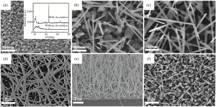
SEM images in Figs. 1(a)-1(c) are the time-dependent morphology evolution of GaAs NWs grown under Ga rate 0.16nm/s and As4 flux × 1.0 10-5 Torr at 580 ℃. After 300s growth, GaAs islands appear around the catalyst droplets on the substrate (Fig. 1(a)). To further illustrate the effects of Au catalyst on GaAs crystallization, we also prepared GaAs on clean fused quartz substrate (without Au) under the same condition. However, as shown in the inset of Fig. 1(a), only very weak crystalline GaAs peak is observed from XRD analysis of the sample grown on clean fused quartz substrate. The NWs grown for 900 s have cylindrical shape, with the wire diameters equal to the catalyst seeds (Fig. 1(b)). For NWs grown for 1800 s, pencil-like morphology was observed, which implies the substantial radial growth of NWs (Fig. 1(c)). Pure axial growth for NWs shown in Fig. 1(b) implies that the reactant adatoms are preferentially dissolved in droplets and deposit at the solid and droplets interfaces. The maximum length of NWs in Fig. 1(b) can reach about 1.2 μm, which is the direct evidence of much longer diffusion length of reactant adatoms for MBE growth than that for CVD growth[6].

The SEM images in Figs. 1(d)-1(f) illustrate GaAs NWs grown on fused quartz substrate for different temperatures. It is observed that NWs are successfully fabricated at various growth temperatures ranging from 480 ℃ (Fig. 1(e)) to 600 ℃ (Fig. 1(f)). In Fig. 1, most NWs end with an Au particle at the top, which implies a typical Au-catalyzed VLS growth mechanism of the NW. However, from 480 ℃ (Fig. 1(e)) to 580 ℃ (Fig. 1(d)) and 600 ℃ (Fig. 1(f)), striking morphological differences can be seen. At 480 ℃, a high density of corn-like NWs is obtained. As shown in Figure S1, corn-like morphology usually takes place in association with the high density of plane defects. Further increasing the temperature to 580 ℃, tapered but straight NWs are observed at the surface of the substrate. At 600 ℃, rod-shaped NWs (with a uniform diameter throughout the entire length) are also observed with much lower density than that grown at 480 ℃ and 580 ℃. At even higher temperature (630 ℃, not shown) nearly no material is present and only large crystallites cover the surface after the same growth duration. Considering the overall morphology and axial growth rate of NWs, we suggest the optimum growth temperature window between 580 ℃ and 600 ℃.

Figure  1.  SEM images of GaAs NWs grown on fused quartz substrates under Ga rate 0.16 nm/s and As4 flux 1.0 × 10-5 Torr.(a)Plane view at 580 ℃, 300 s, the inset shows XRD results of GaAs prepared with and without Au catalyst.(b)Plane view at 580 ℃, 900 s.(c)Plane view at 580 ℃, 1800 s.(d)Plane view at 480 ℃, 3600 s.(e)45ı tilted view at 580 ℃, 3600 s.(f)Plane view at 600 ℃, 3600 s

From the SEM images shown in Fig. 1, one can find most NWs grown by MBE are in a vertical direction relative to the substrate. NWs grown in the horizontal direction parallel to the fused quartz substrate were not observed. Compared with the growth results of NWs grown by MOCVD[21], one can notice that two-dimensional (2D) epitaxy layers form during MBE growth over the full range of experimental times. This can be explained by the diffuse-induced character of MBE technique. Similar phenomena can be observed in GaAs NWs grown on a semiconductor substrate, such as GaAs[12, 17] and Si[18].

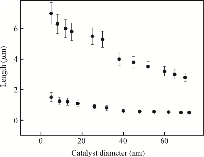
To further understand the growth mechanism of GaAs NWs, the NWs' length and diameter as a function of the seed's diameter for NWs grown at 580 ℃ for 3600 s are summarized in Fig. 2. The statistical data on the lengths of NW show that about 75% NWs are longer than 3 μm, while the rest are shorter than 2 μm. From Fig. 2, one can note that two distinct NWs' lengths are always observed at each point of catalyst diameter. Hence, the expected dependence of NWs' length with catalyst diameter (i.e., L decreasing with increasing d) is not satisfied.

Figure  2.  NW length as a function of the catalyst diameter, NWs grown under Ga rate 0.16 nm/s and As4 flux 1.0 × 10-5 Torr for 3600 s at 580 ℃

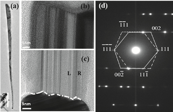
To seek the underlying reasons for the expected inverse dependence of NWs' length with catalyst diameter, closer inspection was performed on two typical NWs with different lengths (the NWs were taken from the sample shown in Fig. 1(e)) by TEM. In Fig. 3(a), we show the TEM image of a longer GaAs NW with about 4 μm in length and about 38 nm in catalyst diameter. From the patterns of selected area electron diffraction (SAED, Fig. 3(d)), the distances between the adjacent lattice planes are found to be 0.28 and 0.33 nm, which are in good agreement with the plane spacing of {200} and {111} families in the ZB structure. The energy dispersive X-ray spectrum (EDX) gives a Ga/As atomic ratio close to 1 : 1 with around 6% O, where this low O content probably comes from the surface oxide layer. (Figure S2.) All these phenomena suggest the NWs are highly crystalline and stoichiometric. Furthermore, SAED analysis of individual NWs confirms that the ZB phase is the dominant crystal structure. This is different from the published results of MBE-grown GaAs NWs on semiconductor substrates, in which the wurtzite (WZ) phase is always reported.

Most importantly, the high-resolution TEM (HRTEM) images viewed in the [¯1¯1¯1] direction show the NW consists of many twin segments of alternating orientation. The segments are separated by twins with the mirror plane parallel to the growth direction. This is evident from the corresponding FFT, Fig. 3(d), where the left (L) and right (R) regions share (¯1¯1¯1) and (111) facets, that is, have a common {111} twin plane. From Fig. 3(d), one can prove that the growth direction of the NW shown in Fig. 3(a) is [11¯2]. Meanwhile, one can find that the longitudinal twins structure runs throughout the whole length of the NW. This characteristic is in good agreement with previous reports on longitudinal defect formation in group IV (Si, Ge)[22, 23] NWs with a [11¯2] growth direction. No transverse plane defects (twin plane and stacking faults) are observed even at the neck region of the NW.

Figure  3.  (a)Overview image of a longer nanowire with a gold particle on its top.The bottom of the wire shown in(b).(b), (c)HRTEM images taken from the top, and bottom of the GaAs nanowire; (d)SAED viewed from the [110] direction

Figure 4 shows TEM results (viewed in the [¯110] direction) of shorter GaAs NWs (0.6 μm in length, 35 nm in catalyst diameter). SAED patterns reveal a typical WZ GaAs NW grown by Au-assisted MBE on semiconductor substrates. Transverse plane defects (stacking faults) are observed over the entire NW, with an approximate frequency as low as 0.03 nm-1.

Figure  4.  (a)HRTEM images taken from the top and bottom of a shorter GaAs NW.(b)Overview image of the GaAs NW, the inset shows SAED viewed from the [110] direction

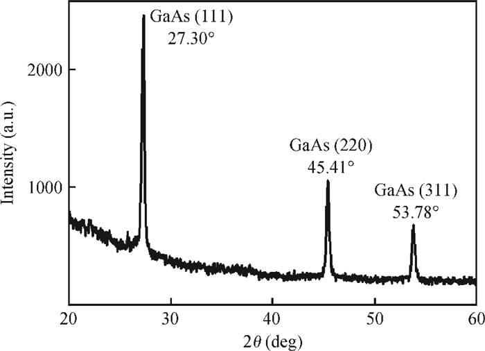
The crystal qualities of GaAs NWs were then studied by XRD as shown in Fig. 5. Based on the XRD spectrum, three diffraction peaks were found at 2θ angle of 27.30, 45.41, and 53.73, which are indexed to GaAs ZB (111), (220), and (311) planes, respectively. No peaks assigned to the hexagonal WZ structure were observed. This also confirms the ZB structure of the NWs, which is consistent with the TEM results. However, in Fig. 5, one could not find any peak corresponding to the small fraction of NWs with WZ structure confirmed by the TEM and SAED characterization, which may be attributed to weak detection ability of conventional XRD.

Figure  5.  XRD diffraction curve obtained from GaAs NWs grown on fused quartz substrate under Ga rate 0.16 nm/s and As4 flux 1.0 × 10-5 Torr for 3600 s at 580 ℃

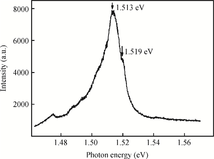
Figure 6 shows the PL spectrum obtained at 4.2 K. Different from reported results for NWs grown on glass substrate by MOCVD[21], strong PL can be observed from the NWs as shown in Fig. 3(a) (ZB structure). The shoulder peak at 1.519eV corresponds closely to the free excitons' emission as reported in bulk GaAs, which further proves the ZB structure of the measured NWs. The dominant (A0-X) peak at 1.513 eV can be related to free excitons bound mainly to neutral acceptors (A0-X). The bound excitons' peak has an FWHM of about 15 meV, which implies high crystal quality of NWs grown on fused-quartz substrate.

Figure  6.  μ-PL spectra recorded from ZB GaAs NW

As the diffuse-induced process, three unique characters can be obtained from the published results of NWs grown by Au-assisted MBE on GaAs substrate[12, 17]: (1) a thin 2-D layer simultaneously formed on the substrate during the NWs' growth. (2) The NW length dependence is found to be close to the inversely proportional relation with catalyst diameter (LA/D). (3) In contrast to their bulk counterparts, NWs grown by MBE often adopt the hexagonal WZ structure and grow in an aligned <0001> direction.

Different from those on GaAs surface as reported by Nitta et al.[24], Ga adatoms incident undergo adsorption and evaporation for MBE growth on the fused quartz substrate. When a clean fused quartz (without Au catalyst) is used as substrate, Ga sticking coefficient is far below unity and a large amount of Ga adatoms desorbed from the SiO2 surface. But when the Au catalyst is introduced, these isolated catalyst droplets can serve as collection centers to accommodate reactive adatoms. 2D epitaxy layer and islands are formed during the process of these reactive adatoms diffusing from substrates to the catalyst droplets. This can be further proved by XRD results, in which more crystallized GaAs can be observed on the amorphous substrate surface with Au catalyst than that on the clean SiO2 surface. Furthermore, the crystallized 2-D layer can serve as a buffer layer for the following growth of NWs.

From Dubrovskii et al.'s diffusion-induced growth model[25, 26] for Ⅲ-Ⅴ NWs, the length of the NW is a function of 1/D (where D is the catalyst diameter). This model takes into account the diffusion of group Ⅲ adatoms from the substrate surface to the NW base and the diffusion of group Ⅲ adatoms along the NW sidewalls to the metallic droplet. However, this model does not consider the effects of crystal structure on the length of NWs grown by MBE. As reported by Glas et al.[27], high group Ⅲ supersaturation is needed to initiate the WZ NW, while low group Ⅲ supersaturation is needed to drive the ZB NW. Accordingly, under the same growth condition (Ga flux, Au diameter), higher axial growth rate can be obtained for ZB NWs as compared with that for WZ NWs. This can explain why the length of ZB NWs is much longer than that of WZ NWs. Furthermore, when the lengths of NWs with the same crystal structure are separately picked up, the inversely proportional relation of NW's lengths with catalyst diameters can still be satisfied. These phenomena further prove the diffusion-induced process of NWs grown by MBE on fused quartz substrate.

Next we discuss the intriguing results of crystal structure of NWs grown on fused quartz substrate. For Au-catalyst MBE growth of NWs on GaAs substrates, group Ⅲ saturation dominates the crystal structure of NWs. WZ NWs were widely reported because the high group Ⅲ saturation is easy to satisfy in Au-assisted MBE growth of GaAs NWs. Meanwhile, ZB GaAs NWs could also be obtained through carefully controlling the diameters of Au catalyst (reducing the group Ⅲ saturation)[28]. To clarify why ZB formed on fused quartz substrates, comparison experiments were also performed on NWs grown on (111) B GaAs substrates. However, under the same growth condition (distribution of Au diameters, growth temperature and Ⅴ/Ⅲ ratio), only WZ structure is observed for NWs grown on GaAs substrate. Obviously, group Ⅲ saturation must not be the major cause of the ZB phase for NWs grown on fused quartz substrate. From Figs. 3(b) and 3(c), one can notice two important phenomena: (1) zigzag-shaped interfaces are formed between Au particles and GaAs NWs (Fig.3(b)), (2) lamellar {111} twins extend through the length of GaAs NWs in [11¯2] growth direction. Considering these, one can deduce that GaAs nucleate at the catalyst/substrate interfaces to form the saw-teeth shaped interface at the very beginning of NWs growth. As discussed previously in Ref. [29], nucleation at the liquid-solid interface favors the ZB phase formation. The promotion of liquid-solid interface nucleation may be attributed to the interface energy change between catalyst, nucleated GaAs and the amorphous substrate. To understand why GaAs NWs grown in [11¯2] direction rather than [110] or [100] directions as reported by Shtrikman et al.[28], one can consider the overall system energy for GaAs NWs grown in different directions. As reported in Ref. [30], the sidewall of the [11¯2] NWs is wrapped with {111} and {311} side facets. These side facets have lower surface energy than {110} facets associated with the [110] NWs. For longer NWs (considering the high axial growth rate of [11¯2] NWs), lower surface energy helps to maintain lowest overall system energy for GaAs NWs.

It is of interest to note that the PL results show high-intensity exciton emission for NWs grown in [11¯2] direction, although the {111} twins' defects extended through the length of NWs. This implies that this longitudinal planar defect is not the effective recombination center. Furthermore, we didn't find a carbon-impurity related PL peak as reported by Joyce et al.[8], which proves the advantages (ultra-high vacuum, ultra-purity source) of MBE technique.

GaAs NWs were grown on fused quartz substrate by Au-assisted MBE. A typical diffuse-induced dominant process can be observed from the NWs grown by MBE as compared with the reported results of NWs grown on glass substrate by MOCVD. TEM results show that ZB crystal structure and [11¯2] growth direction are dominant for NWs grown on fused quartz substrate by Au-assisted MBE. Lamellar {111} twins extended through the GaAs NWs with [11¯2] growth direction. Although there are longitudinal planar defects extending through NWs, the strong narrow FWHM implies high crystal quality of NWs grown on fused quartz substrate.


[1]
Doh Y J, van Dam J A, Roest A L, et al. Tunable supercurrent through semiconductor nanowires. Science, 2005, 309(5732):272 doi: 10.1126/science.1113523
[2]
Glas F, Harmand J C, Patriarche G. Why does wurtzite form in nanowires of Ⅲ-Ⅴ zinc blende semiconductors. Phys Rev Lett, 2007, 99(14):146101 doi: 10.1103/PhysRevLett.99.146101
[3]
Lu W, Lieber C M. Nanoelectronics from the bottom up. Nat Mater, 2007, 6(11):841 doi: 10.1038/nmat2028
[4]
Qian F, Gradecak S, Li Y, et al. Core/multishell nanowire heterostructures as multicolor, high-efficiency light-emitting diodes. Nano Lett, 2005, 5(11):2287 doi: 10.1021/nl051689e
[5]
Raj G, Pardeshi H, Pati K, et al. 2DEG charge density based drain current model for nano-scale AlInGaN/AlN/GaN HEMT devices. Journal of Semiconductors, 2013, 34(4):044002 doi: 10.1088/1674-4926/34/4/044002
[6]
Bao X Y, Soci C, Susac D, et al. Heteroepitaxial growth of vertical GaAs nanowires on Si (111) substrates by metal-organic chemical vapor deposition. Nano Lett, 2008, 8(11):3755 doi: 10.1021/nl802062y
[7]
Xu H Y, Guo Y N, Sun W, et al. Quantitative study of GaAs nanowires catalyzed by Au film of different thicknesses. Nanoscale Res Lett, 2012, 7(1):1 doi: 10.1186/1556-276X-7-1
[8]
Joyce H J, Gao Q, Tan H H, et al. High purity GaAs nanowires free of planar defects:growth and characterization. Adv Funct Mater, 2008, 18(23):3794 doi: 10.1002/adfm.v18:23
[9]
Lugani L, Ercolani D, Sorba L, et al. Modeling of InAs-InSb nanowires grown by Au-assisted chemical beam epitaxy. Nanotechnology, 2012, 23(9):095602 doi: 10.1088/0957-4484/23/9/095602
[10]
Ercolani D, Rossi F, Li A, et al. InAs/InSb nanowire heterostructures grown by chemical beam epitaxy. Nanotechnology, 2009, 20(50):505605 doi: 10.1088/0957-4484/20/50/505605
[11]
Plissard S, Dick K A, Larrieu G, et al. Gold-free growth of GaAs nanowires on silicon:arrays and polytypism. Nanotechnology, 2010, 21(38):385602 doi: 10.1088/0957-4484/21/38/385602
[12]
Tchernycheva M, Harmand J C, Patriarche G, et al. Temperature conditions for GaAs nanowire formation by Au-assisted molecular beam epitaxy. Nanotechnology, 2006, 17(16):4025 doi: 10.1088/0957-4484/17/16/005
[13]
Duan X, Lieber C M. General synthesis of compound semiconductor nanowires. Adv Mater, 2000, 12(4):298 doi: 10.1002/(ISSN)1521-4095
[14]
Shao Y, Nie T, Jiang Z, et al. Behavior of Au-Si droplets in Si (001) at high temperatures. Appl Phys Lett, 2012, 101(5):053104 doi: 10.1063/1.4739413
[15]
Wagner R, Ellis W. Vapor-liquid-solid mechanism of single crystal growth. Appl Phys Lett, 1964, 4(5):89 doi: 10.1063/1.1753975
[16]
Kayes B M, Atwater H A, Lewis N S. Comparison of the device physics principles of planar and radial p-n junction nanorod solar cells. J Appl Phys, 2005, 97(11):114302 doi: 10.1063/1.1901835
[17]
Plante M C, Lapierre R R. Control of GaAs nanowire morphology and crystal structure. Nanotechnology, 2008, 9(49):495603
[18]
Kang J H, Gao Q, Joyce H J, et al. Novel growth and properties of GaAs nanowires on Si substrates. Nanotechnology, 2010, 21(3):035604 doi: 10.1088/0957-4484/21/3/035604
[19]
Akinlami J O, Ashamu A O. Optical properties of gallium arsenide GaAs. Journal of Semiconductors, 2013, 34(3):032002 doi: 10.1088/1674-4926/34/3/032002
[20]
Wen L, Li X, Zhao Z, et al. Theoretical consideration of Ⅲ-Ⅴ nanowire/Si triple-junction solar cells. Nanotechnology, 2012, 23(50):505202 doi: 10.1088/0957-4484/23/50/505202
[21]
Dhaka V, Haggren T, Jussila H, et al. High quality GaAs nanowires grown on glass substrates. Nano Lett, 2012, 12(4):1912 doi: 10.1021/nl204314z
[22]
Geaney H, Dickinson C, Weng W, et al. Role of defects and growth directions in the formation of periodically twinned and kinked unseeded germanium nanowires. Cryst Growth Des, 2011, 11(7):3266 doi: 10.1021/cg200510y
[23]
Davidson F M, Lee D C, Fanfair D D, et al. Lamellar twinning in semiconductor nanowires. J Phys Chem C, 2007, 111(7):2929 doi: 10.1021/jp0672205
[24]
Nitta Y, Ohta J, Mitsunaga K, et al. GaAs/AlGaAs optical interconnection chip for neural network. Jpn J Appl Phys Lett, 1989, 28(11):L2101 https://confit.atlas.jp/guide/organizer/ssdm/ssdm1989/subject/S-C-1/search;jsessionid=76DDD57608627B90100FAFF5D46460D9?eventCode=ssdm1989&code=S-C-1
[25]
Dubrovskii V, Sibirev N, Cirlin G, et al. Theoretical analysis of the vapor-liquid-solid mechanism of nanowire growth during molecular beam epitaxy. Phys Rev E, 2006, 73(2):021603 https://www.ncbi.nlm.nih.gov/pubmed/16605346
[26]
Dubrovskii V, Cirlin G, Soshnikov I, et al. Diffusion-induced growth of GaAs nanowhiskers during molecular beam epitaxy:theory and experiment. Phys Rev B, 2005, 71(20):205325 doi: 10.1103/PhysRevB.71.205325
[27]
Glas F, Harmand J C, Patriarche G. Why does wurtzite form in nanowires of Ⅲ-Ⅴ zinc blende semiconductors. Phys Rev Lett, 2007, 99(14):146101 doi: 10.1103/PhysRevLett.99.146101
[28]
Shtrikman H, Popovitz-Biro R, Kretinin A, et al. Stacking-faults-free zinc blende GaAs nanowires. Nano Lett, 2008, 9(1):215 https://www.ncbi.nlm.nih.gov/pubmed/19093840
[29]
Cirlin G, Dubrovskii V, Samsonenko Y B, et al. Self-catalyzed, pure zinc-blende GaAs nanowires grown on Si (111) by molecular beam epitaxy. Phys Rev B, 2010, 82(3):035302 doi: 10.1103/PhysRevB.82.035302
[30]
Li C P, Lee C S, Ma X L, et al. Growth direction and cross-sectional study of silicon nanowires. Adv Mater, 2003, 15(7/8):607 http://repository.ust.hk/ir/Record/1783.1-22648
Fig. 1.  SEM images of GaAs NWs grown on fused quartz substrates under Ga rate 0.16 nm/s and As4 flux 1.0 × 10-5 Torr.(a)Plane view at 580 ℃, 300 s, the inset shows XRD results of GaAs prepared with and without Au catalyst.(b)Plane view at 580 ℃, 900 s.(c)Plane view at 580 ℃, 1800 s.(d)Plane view at 480 ℃, 3600 s.(e)45ı tilted view at 580 ℃, 3600 s.(f)Plane view at 600 ℃, 3600 s

Fig. 2.  NW length as a function of the catalyst diameter, NWs grown under Ga rate 0.16 nm/s and As4 flux 1.0 × 10-5 Torr for 3600 s at 580 ℃

Fig. 3.  (a)Overview image of a longer nanowire with a gold particle on its top.The bottom of the wire shown in(b).(b), (c)HRTEM images taken from the top, and bottom of the GaAs nanowire; (d)SAED viewed from the [110] direction

Fig. 4.  (a)HRTEM images taken from the top and bottom of a shorter GaAs NW.(b)Overview image of the GaAs NW, the inset shows SAED viewed from the [110] direction

Fig. 5.  XRD diffraction curve obtained from GaAs NWs grown on fused quartz substrate under Ga rate 0.16 nm/s and As4 flux 1.0 × 10-5 Torr for 3600 s at 580 ℃

Fig. 6.  μ-PL spectra recorded from ZB GaAs NW

[1]
Doh Y J, van Dam J A, Roest A L, et al. Tunable supercurrent through semiconductor nanowires. Science, 2005, 309(5732):272 doi: 10.1126/science.1113523
[2]
Glas F, Harmand J C, Patriarche G. Why does wurtzite form in nanowires of Ⅲ-Ⅴ zinc blende semiconductors. Phys Rev Lett, 2007, 99(14):146101 doi: 10.1103/PhysRevLett.99.146101
[3]
Lu W, Lieber C M. Nanoelectronics from the bottom up. Nat Mater, 2007, 6(11):841 doi: 10.1038/nmat2028
[4]
Qian F, Gradecak S, Li Y, et al. Core/multishell nanowire heterostructures as multicolor, high-efficiency light-emitting diodes. Nano Lett, 2005, 5(11):2287 doi: 10.1021/nl051689e
[5]
Raj G, Pardeshi H, Pati K, et al. 2DEG charge density based drain current model for nano-scale AlInGaN/AlN/GaN HEMT devices. Journal of Semiconductors, 2013, 34(4):044002 doi: 10.1088/1674-4926/34/4/044002
[6]
Bao X Y, Soci C, Susac D, et al. Heteroepitaxial growth of vertical GaAs nanowires on Si (111) substrates by metal-organic chemical vapor deposition. Nano Lett, 2008, 8(11):3755 doi: 10.1021/nl802062y
[7]
Xu H Y, Guo Y N, Sun W, et al. Quantitative study of GaAs nanowires catalyzed by Au film of different thicknesses. Nanoscale Res Lett, 2012, 7(1):1 doi: 10.1186/1556-276X-7-1
[8]
Joyce H J, Gao Q, Tan H H, et al. High purity GaAs nanowires free of planar defects:growth and characterization. Adv Funct Mater, 2008, 18(23):3794 doi: 10.1002/adfm.v18:23
[9]
Lugani L, Ercolani D, Sorba L, et al. Modeling of InAs-InSb nanowires grown by Au-assisted chemical beam epitaxy. Nanotechnology, 2012, 23(9):095602 doi: 10.1088/0957-4484/23/9/095602
[10]
Ercolani D, Rossi F, Li A, et al. InAs/InSb nanowire heterostructures grown by chemical beam epitaxy. Nanotechnology, 2009, 20(50):505605 doi: 10.1088/0957-4484/20/50/505605
[11]
Plissard S, Dick K A, Larrieu G, et al. Gold-free growth of GaAs nanowires on silicon:arrays and polytypism. Nanotechnology, 2010, 21(38):385602 doi: 10.1088/0957-4484/21/38/385602
[12]
Tchernycheva M, Harmand J C, Patriarche G, et al. Temperature conditions for GaAs nanowire formation by Au-assisted molecular beam epitaxy. Nanotechnology, 2006, 17(16):4025 doi: 10.1088/0957-4484/17/16/005
[13]
Duan X, Lieber C M. General synthesis of compound semiconductor nanowires. Adv Mater, 2000, 12(4):298 doi: 10.1002/(ISSN)1521-4095
[14]
Shao Y, Nie T, Jiang Z, et al. Behavior of Au-Si droplets in Si (001) at high temperatures. Appl Phys Lett, 2012, 101(5):053104 doi: 10.1063/1.4739413
[15]
Wagner R, Ellis W. Vapor-liquid-solid mechanism of single crystal growth. Appl Phys Lett, 1964, 4(5):89 doi: 10.1063/1.1753975
[16]
Kayes B M, Atwater H A, Lewis N S. Comparison of the device physics principles of planar and radial p-n junction nanorod solar cells. J Appl Phys, 2005, 97(11):114302 doi: 10.1063/1.1901835
[17]
Plante M C, Lapierre R R. Control of GaAs nanowire morphology and crystal structure. Nanotechnology, 2008, 9(49):495603
[18]
Kang J H, Gao Q, Joyce H J, et al. Novel growth and properties of GaAs nanowires on Si substrates. Nanotechnology, 2010, 21(3):035604 doi: 10.1088/0957-4484/21/3/035604
[19]
Akinlami J O, Ashamu A O. Optical properties of gallium arsenide GaAs. Journal of Semiconductors, 2013, 34(3):032002 doi: 10.1088/1674-4926/34/3/032002
[20]
Wen L, Li X, Zhao Z, et al. Theoretical consideration of Ⅲ-Ⅴ nanowire/Si triple-junction solar cells. Nanotechnology, 2012, 23(50):505202 doi: 10.1088/0957-4484/23/50/505202
[21]
Dhaka V, Haggren T, Jussila H, et al. High quality GaAs nanowires grown on glass substrates. Nano Lett, 2012, 12(4):1912 doi: 10.1021/nl204314z
[22]
Geaney H, Dickinson C, Weng W, et al. Role of defects and growth directions in the formation of periodically twinned and kinked unseeded germanium nanowires. Cryst Growth Des, 2011, 11(7):3266 doi: 10.1021/cg200510y
[23]
Davidson F M, Lee D C, Fanfair D D, et al. Lamellar twinning in semiconductor nanowires. J Phys Chem C, 2007, 111(7):2929 doi: 10.1021/jp0672205
[24]
Nitta Y, Ohta J, Mitsunaga K, et al. GaAs/AlGaAs optical interconnection chip for neural network. Jpn J Appl Phys Lett, 1989, 28(11):L2101 https://confit.atlas.jp/guide/organizer/ssdm/ssdm1989/subject/S-C-1/search;jsessionid=76DDD57608627B90100FAFF5D46460D9?eventCode=ssdm1989&code=S-C-1
[25]
Dubrovskii V, Sibirev N, Cirlin G, et al. Theoretical analysis of the vapor-liquid-solid mechanism of nanowire growth during molecular beam epitaxy. Phys Rev E, 2006, 73(2):021603 https://www.ncbi.nlm.nih.gov/pubmed/16605346
[26]
Dubrovskii V, Cirlin G, Soshnikov I, et al. Diffusion-induced growth of GaAs nanowhiskers during molecular beam epitaxy:theory and experiment. Phys Rev B, 2005, 71(20):205325 doi: 10.1103/PhysRevB.71.205325
[27]
Glas F, Harmand J C, Patriarche G. Why does wurtzite form in nanowires of Ⅲ-Ⅴ zinc blende semiconductors. Phys Rev Lett, 2007, 99(14):146101 doi: 10.1103/PhysRevLett.99.146101
[28]
Shtrikman H, Popovitz-Biro R, Kretinin A, et al. Stacking-faults-free zinc blende GaAs nanowires. Nano Lett, 2008, 9(1):215 https://www.ncbi.nlm.nih.gov/pubmed/19093840
[29]
Cirlin G, Dubrovskii V, Samsonenko Y B, et al. Self-catalyzed, pure zinc-blende GaAs nanowires grown on Si (111) by molecular beam epitaxy. Phys Rev B, 2010, 82(3):035302 doi: 10.1103/PhysRevB.82.035302
[30]
Li C P, Lee C S, Ma X L, et al. Growth direction and cross-sectional study of silicon nanowires. Adv Mater, 2003, 15(7/8):607 http://repository.ust.hk/ir/Record/1783.1-22648
1

Effect of nitrogen incorporation and surface passivation on photoluminescence properties of InAs-based nanowires

Ratmir Ustimenko, Danila Karaulov, Maxim Vinnichenko, Ilya Norvatov, Andrey Kaveev, et al.

Journal of Semiconductors. doi: 10.1088/1674-4926/25030041

2

The scanning tunneling microscopy and spectroscopy of GaSb1–xBix films of a few-nanometer thickness grown by molecular beam epitaxy

Fangxing Zha, Qiuying Zhang, Haoguang Dai, Xiaolei Zhang, Li Yue, et al.

Journal of Semiconductors, 2021, 42(9): 092101. doi: 10.1088/1674-4926/42/9/092101

3

Research on the photoluminescence of spectral broadening by rapid thermal annealing on InAs/GaAs quantum dots

Dandan Ning, Yanan Chen, Xinkun Li, Dechun Liang, Shufang Ma, et al.

Journal of Semiconductors, 2020, 41(12): 122101. doi: 10.1088/1674-4926/41/12/122101

4

Controllable growth of GeSi nanostructures by molecular beam epitaxy

Yingjie Ma, Tong Zhou, Zhenyang Zhong, Zuimin Jiang

Journal of Semiconductors, 2018, 39(6): 061004. doi: 10.1088/1674-4926/39/6/061004

5

Manipulation of morphology and structure of the top of GaAs nanowires grown by molecular-beam epitaxy

Lixia Li, Dong Pan, Xuezhe Yu, Hyok So, Jianhua Zhao, et al.

Journal of Semiconductors, 2017, 38(10): 103001. doi: 10.1088/1674-4926/38/10/103001

6

Analysis of morphological, structural and electrical properties of annealed TiO2 nanowires deposited by GLAD technique

B. Shougaijam, R. Swain, C. Ngangbam, T.R. Lenka

Journal of Semiconductors, 2017, 38(5): 053001. doi: 10.1088/1674-4926/38/5/053001

7

A GaAs/GaInP dual junction solar cell grown by molecular beam epitaxy

Pan Dai, Shulong Lu, Lian Ji, Wei He, Lifeng Bian, et al.

Journal of Semiconductors, 2013, 34(10): 104006. doi: 10.1088/1674-4926/34/10/104006

8

GaAs-based long-wavelength InAs bilayer quantum dots grown by molecular beam epitaxy

Zhu Yan, Li Mifeng, He Jifang, Yu Ying, Ni Haiqiao, et al.

Journal of Semiconductors, 2011, 32(8): 083001. doi: 10.1088/1674-4926/32/8/083001

9

Size-independent growth of pure zinc blende GaAs nanowires

Ye Xian, Huang Hui, Guo Jingwei, Ren Xiaomin, Huang Yongqing, et al.

Journal of Semiconductors, 2010, 31(7): 073001. doi: 10.1088/1674-4926/31/7/073001

10

Visible photoluminescence of porous silicon covered with an HfON dielectric layer

Jiang Ran, Zhang Yan

Journal of Semiconductors, 2009, 30(8): 082003. doi: 10.1088/1674-4926/30/8/082003

11

Low-Temperature Growth and Photoluminescence of SnO2 Nanowires

Wang Bing, Xu Ping, Yang Guowei

Journal of Semiconductors, 2008, 29(8): 1469-1474.

12

The Origin of Multi-Peak Structures Observed in Photoluminescence Spectra of InAs/GaAs Quantum Dots

Liang Zhimei, Wu Ju, Jin Peng, Lü Xueqin, Wang Zhanguo, et al.

Journal of Semiconductors, 2008, 29(11): 2121-2124.

13

Synthesis and Photoluminescence Properties of Dy-Doped ZnO Nanocrystals

Zhang Linli, Guo Changxin

Chinese Journal of Semiconductors , 2007, 28(6): 887-892.

14

High Resistivity Step-Graded InAIAs/GaAs Metamorphic Buffer Layers Grown by Molecular Beam Epitaxy in Low Temperature

Gao Hongling, Wang Baoqiang, Zhu Zhanping, Li Chengji, Duan Ruifei, et al.

Chinese Journal of Semiconductors , 2007, 28(S1): 200-203.

15

Growth of AlGaAs on GaAs (110) Surface by Molecular Beam Epitaxy

Liu Linsheng, Wang Wenxin, Liu Su, Zhao Hongming, Liu Baoli, et al.

Chinese Journal of Semiconductors , 2007, 28(9): 1411-1414.

16

Photoluminescence of nc-Si/SiN Superlattices Embeddedin Optical Microcavities

Chen San, Qian Bo, Chen Kunji, Cen Zhanhong, Liu Yansong, et al.

Chinese Journal of Semiconductors , 2006, 27(S1): 25-28.

17

An Evanescent Coupling Approach for Optical Characterization of ZnO Nanowires

Yang Qing, Lou Jingyi, Yang Deren, Tong Limin

Chinese Journal of Semiconductors , 2006, 27(3): 425-428.

18

Photoluminescence During the Crystallization of a-Si∶H/SiO2 Multilayers

Ma Zhongyuan, Han Peigao, Li Wei, Chen San, Qian Bo, et al.

Chinese Journal of Semiconductors , 2006, 27(S1): 76-79.

19

1.3μm InGaAs/InAs/GaAs Self-Assembled Quantum Dot Laser Diode Grown by Molecular Beam Epitaxy

Niu Zhichuan, Ni Haiqiao, Fang Zhidan, Gong Zheng, Zhang Shiyong, et al.

Chinese Journal of Semiconductors , 2006, 27(3): 482-488.

20

Resonant Raman Scattering and Photoluminescence Emissions from Above Bandgap Levels in Dilute GaAsN Alloys

Tan P H, Luo X D, Ge W K, Xu Z Y, Zhang Y, et al.

Chinese Journal of Semiconductors , 2006, 27(3): 397-402.

  • Search

    Advanced Search >>

    GET CITATION

    Yufeng Zhao, Xinhua Li, Wenbo Wang, Bukang Zhou, Huahua Duan, Tongfei Shi, Xuesong Zeng, Ning Li, Yuqi Wang. Growth and properties of GaAs nanowires on fused quartz substrate[J]. Journal of Semiconductors, 2014, 35(9): 093002. doi: 10.1088/1674-4926/35/9/093002
    Y F Zhao, X H Li, W B Wang, B K Zhou, H H Duan, T F Shi, X S Zeng, N Li, Y Q Wang. Growth and properties of GaAs nanowires on fused quartz substrate[J]. J. Semicond., 2014, 35(9): 093002. doi: 10.1088/1674-4926/35/9/093002.
    shu

    Export: BibTex EndNote

    Article Metrics

    Article views: 3133 Times PDF downloads: 34 Times Cited by: 0 Times

    History

    Received: 25 February 2014 Revised: 17 March 2014 Online: Published: 01 September 2014

    Catalog

      Email This Article

      User name:
      Email:*请输入正确邮箱
      Code:*验证码错误
      Yufeng Zhao, Xinhua Li, Wenbo Wang, Bukang Zhou, Huahua Duan, Tongfei Shi, Xuesong Zeng, Ning Li, Yuqi Wang. Growth and properties of GaAs nanowires on fused quartz substrate[J]. Journal of Semiconductors, 2014, 35(9): 093002. doi: 10.1088/1674-4926/35/9/093002 ****Y F Zhao, X H Li, W B Wang, B K Zhou, H H Duan, T F Shi, X S Zeng, N Li, Y Q Wang. Growth and properties of GaAs nanowires on fused quartz substrate[J]. J. Semicond., 2014, 35(9): 093002. doi: 10.1088/1674-4926/35/9/093002.
      Citation:
      Yufeng Zhao, Xinhua Li, Wenbo Wang, Bukang Zhou, Huahua Duan, Tongfei Shi, Xuesong Zeng, Ning Li, Yuqi Wang. Growth and properties of GaAs nanowires on fused quartz substrate[J]. Journal of Semiconductors, 2014, 35(9): 093002. doi: 10.1088/1674-4926/35/9/093002 ****
      Y F Zhao, X H Li, W B Wang, B K Zhou, H H Duan, T F Shi, X S Zeng, N Li, Y Q Wang. Growth and properties of GaAs nanowires on fused quartz substrate[J]. J. Semicond., 2014, 35(9): 093002. doi: 10.1088/1674-4926/35/9/093002.

      Growth and properties of GaAs nanowires on fused quartz substrate

      DOI: 10.1088/1674-4926/35/9/093002
      Funds:

      Project supported by the National Natural Science Foundation of China (Nos. 11104271, 11179042)

      the National Natural Science Foundation of China 11104271

      the National Natural Science Foundation of China 11179042

      More Information
      • Corresponding author: Li Xinhua, Email:xinhuali@issp.ac.cn
      • Received Date: 2014-02-25
      • Revised Date: 2014-03-17
      • Published Date: 2014-09-01

      Catalog

        /

        DownLoad:  Full-Size Img  PowerPoint
        Return
        Return